
CHAPTER 2, CIRCULATORRY SYSTEM, BRANCHES OF THE AORTA
CIRCULATORRY SYSTEM, BRANCHES OF THE AORTA
[ ইংরেজীতে প্রকাশিত করা হল কিছু কিছু বাংলাদেশী বংশোদ্ভূত বিদেশে অবস্থানরত ছাত্র পাঠকেরা যারা বাংলা ভাষা পড়তে পারেনা, তাদের অনুরোধে, দুখিত ]
Dear viewers,
This is a very simple and preliminary chapter where it has been shown that what is the function of the Aorta and how it is branched primarily.
This might be some useful for the students who who has aim to enter medical line.
To know in detail please open the link below.
AN INTEROR VIEW OF THE HEART WITH ANIMATION
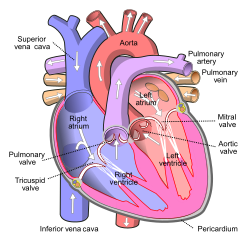
FIGURE 1
This figure shows the Heart action with animation
Look this video
Look the branches of the Aorta
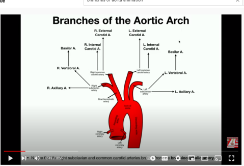
FIGURE 2
See the link
https://www.youtube.com/watch?v=GTk1PCge93A&t=81s
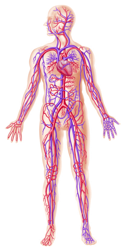
FIGURE 3
https://www.youtube.com/watch?v=SwHjwO7BnsI&t=4s
Dated- 10/30/2021
CHAPTER 1, HOW HEART FUNCTIONS.
[ ইংরেজীতে প্রকাশিত করা হল কিছু কিছু বাংলাদেশী বংশোদ্ভূত বিদেশে অবস্থানরত ছাত্র পাঠকেরা যারা বাংলা ভাষা পড়তে পারেনা, তাদের অনুরোধে, দুখিত ]
HOW HEART FUNCTIONS.
Dear viewers,
Here are some photos and videos of Heart and circulatory system along with link. It can help some students who intend to enter medical school and also those who are interested to have some very primary knowledge about heart and circulatory system.
.Please open the link to know clearly.
HEART SURFACE ANATOMY
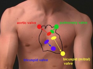
FIGURE 1
This is a body surface marking showing boundary of the heart as located inside the body.
DESCIPTION OF INTERIOR HEART FUCTION with animation.
See this link
https://www.youtube.com/watch?v=BEWjOCVEN7M
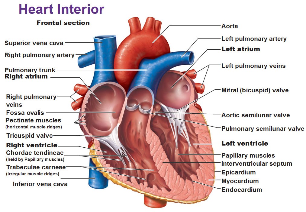
FIGURE 2
THIS PHOTO SHOWS THE CIRCULATION OF HEART
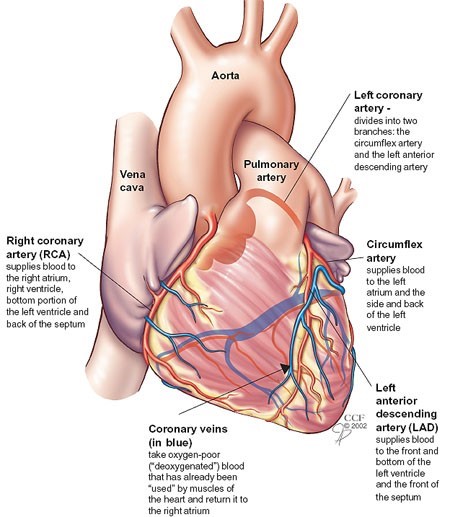
FIGURE 3
This figure shows how the Heart itself receives blood supply.
See this video link.
https://chkdr02.files.wordpress.com/2014/09/37b.jpg
AN EXTERNAL VIEW OF THE HEART
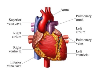
FIGURE 4
This figure shows just the external view of the heart.
https://chkdr02.files.wordpress.com/2014/09/29.jpg
HEART BLOCK BY ARTERIOSCLEROSIS
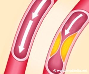
FIGURE 5
Please look how our Coronary vessels, that supply blood to our Heart, are gradually blocking by deposition of Cholesterol or fat.
AN INTEROR VIEW OF THE HEART WITH ANIMATION

FIGURE 6
This figure shows the Heart action with animation
Look this video
HEART CONTRACTION WITH ANIMATION
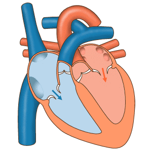
FIGURE 7
This figure also shows the heart function with animation
See this video
CARDIAC CONDUCTION WITH ANIMATION AND EKG
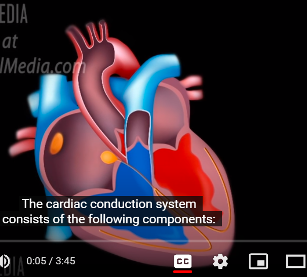
FIGURE 8
This figure shows the electrictric impulse conducts in the Heart
See the video
https://www.youtube.com/watch?v=RYZ4daFwMa8&list=LLwbBmvre5US6kA7XCASuXyQ&index=2
BODY CIRCULATION
SHOWING WHOLE BODY CIRCULATION
Please look how our body is encircled by blood vessel network.
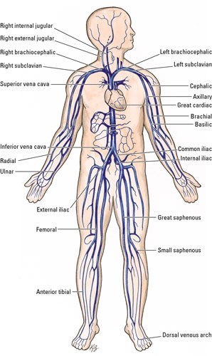
FIGURE 9
EKG INTERPRETATION
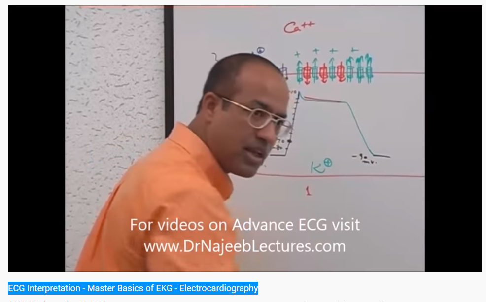
10
FIGURE 10
Please listen the interpretation of ECG/EKG.
ECG Interpretation – Master Basics of EKG – Electrocardiography
CARDIAC DEPOLARIZATION AND REPOLARIZATION AND ACTION POTENTIAL
See this link
https://www.youtube.com/watch?v=IOvmhrmN3p0&list=LLwbBmvre5US6kA7XCASuXyQ
FIGURE 11
ACTION POTEN TIAL, DEPOLARIZATION,REPOLARIZATION, REFRACTORY CONDITION.
What is Action potential?
Action potential is a very short time, sudden and temporary event of Neuron. In this event a sudden rise (Depolarization) followed by sudden fall (Repolarization) of electric membrane potential upon Neuron membrane happens. This very short time sudden rise and fall of electric membrane potential is called the Action potential. This is also called Spike. (2, 6,7, 8)
How does the Neuron produce Action potential?
While cell remains at resting condition, it keeps at-70 m.volt charge on inside wall and positive charge on outside wall. This condition is called the ‘Polarized” condition of cell. At this time, there remains a lot of Sodium ion (Na+) outside the cell wall (See figure-2 for Na, figure-9 voltmeter)
To produce Action potential, it is very essential to hold -70 millivolt charge inside cell wall or it will be unable to produce an Action potential. This is called the resting condition of cell. The cell holds this resting condition through Na+/K+ ion pump mechanism.
At this time, there remains negative charged protein, Nucleic acid, and positive charged potassium ion (k+). (See figure-3, for K)
When Dendrite receives an enough strong signaling charge, it works as a stimulant causing a thrust upon cell. Then the thrust of stimulating action opens the Na ion channel at Axon Hillock. At this time, a lot of Na+ ion enter inside the cell wall and raise the -70 millivolt charge towards positive. This is called Depolarization.
If the signaling charge becomes so strong that it can raise the charge up to threshold level, (-55) then the Neuron fires and a lot of Na+ ion enter inside the cell wall raising the charge as high as up to +40 m.volt that is also called spike.
Now the cell controls and prevents the charge rising farther and fells lower.
Such a very short-time event of sudden rising and falling of change upon a Neuron membrane is called the Action potential. It takes 2 milliseconds (0.0002 sec.) to happen.
How does the cell prevent rising the electric potential above and will lower?
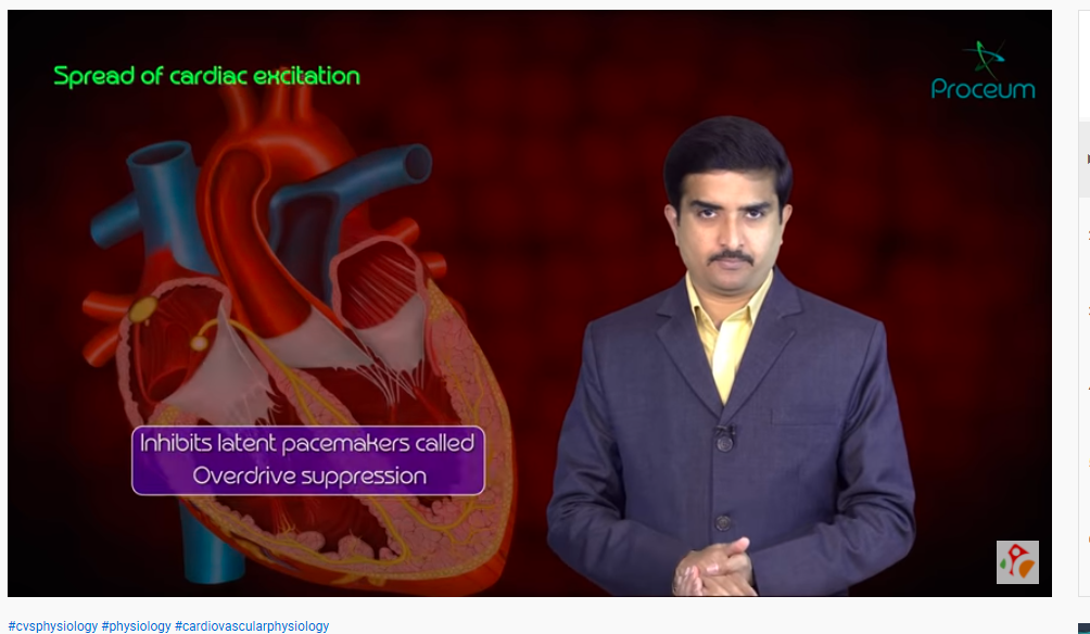
FIGURE- 12
https://www.youtube.com/watch?v=-13IfTvjZRI&list=LLwbBmvre5US6kA7XCASuXyQ
Action potential of cardiac muscle and SA Node – Usmle step 1 CVS Physiology
Date-10/23/202
Blog link-
