
CHAPTER 33, WHAT ARE THE TUNICS, SEGMENTS AND ACCOMODATION OF EYE BALL? WHAT ARE THE STRUCTURE OF RETINA,CHOROID, SCLERA, LENS AND CORNEA?
[ইংরেজীতে প্রকাশিত করা হল কিছু কিছু বাংলাদেশী বংশোদ্ভূত বিদেশে অবস্থানরত ছাত্র পাঠকেরা যারা বাংলা ভাষা পড়তে পারেনা, তাদের অনুরোধে, দুখিত ]
Dear viewers,
It is written as a primary knowledge for the students who are interested in studying in edical sciences. Today we are going to discuss about the tunics of the eye ball, structure of Retina, Sclera and cornea.
LAYERS OF SCLERA TEXT
The sclera has four layers, from the outside to the inside:
- Episclera, clear, thin tissue resting on top of the whites of your eyeballs.Stroma, made up of fibroblasts and collagen fibers, blending into the episclera.
- Lamina fusca, a transitional layer between the sclera and the choroid and ciliary body outer layers.
- Endothelium, the basal, or innermost layer of the sclera.
SOURCE OF TEXT-
https://my.clevelandclinic.org/health/body/22088-sclera
Special Senses | Eye Anatomy, Eye Tunics
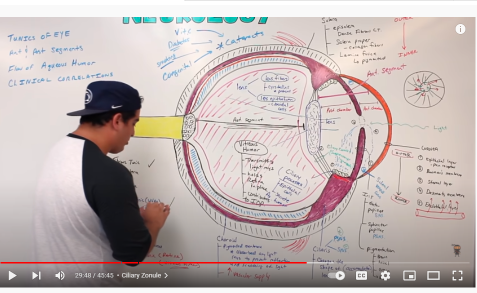
FIGURE-1
SOURCE OF YOU TUBE LINK- https://www.youtube.com/watch?v=Hclc7lL_Oyw
WHAT IS CHOROID? TEXT
What is choroid?
A thin layer of tissue that is part of the middle layer of the wall of the eye, between the sclera (white outer layer of the eye) and the retina (the inner layer of nerve tissue at the back of the eye). The choriod is filled with blood vessels that bring oxygen and nutrients to the eye.
SOURCE OF THE TEXT LINK– https://www.cancer.gov/publications/dictionaries/cancer-terms/def/choroid
STRUCTURE OF RETINA TEXT
FIGURE 2
SOURCE OF FIGURE TEXT- https://webvision.med.utah.edu/book/part-i-foundations/simple-anatomy-of-the-retina/
STRUCTURE OF RETINA TEXT
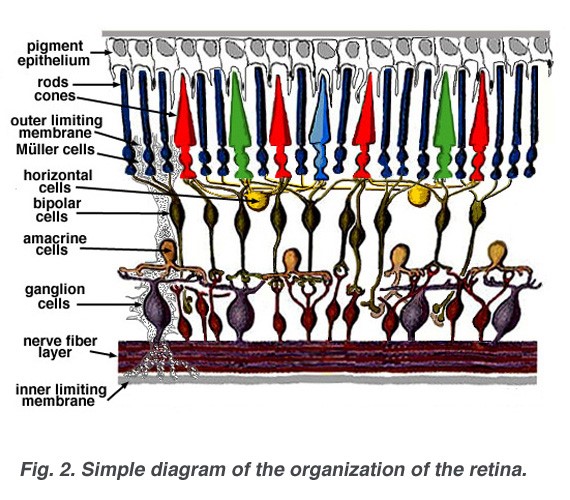
FIGURE 2
SOURCE OF FIGURE TEXT- https://webvision.med.utah.edu/book/part-i-foundations/simple-anatomy-of-the-retina/
STRUCTURE OR LAYERS OF CORNEA TEXT
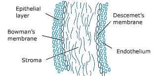
FIGURE- 3
SOURCE OF THE TEXT LINK- https://en.excimerclinic.ru/press/rogovica/
LENS STRUCTURE YOU TUBE
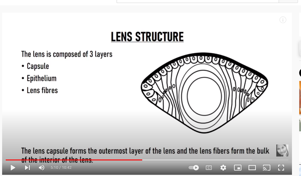
FIGURE 4- https://www.youtube.com/watch?v=kOUYeCWN4g8 SOURCE OF
DATE- date- 5/22/2022
BLOG LINK-
CHAPTER 32, WHAT IS EYE LID AND EYE LASHES? WHAT IS CONJUCTIVA ? WHAT IS NASOLACRIMAL APPARATUS?
[ ইংরেজীতে প্রকাশিত করা হল কিছু কিছু বাংলাদেশী বংশোদ্ভূত বিদেশে অবস্থানরত ছাত্র পাঠকেরা যারা বাংলা ভাষা পড়তে পারেনা, তাদের অনুরোধে, দুখিত ]
Dear viewers,
It is written as a primary knowledge for the students who are interested in studying medical sciences.
It may be somewhat useful for them. Today we are going to discuss about NASOLACRIMAL APPARATUS
Orbit and Eye – Conjunctiva YOU TUBE
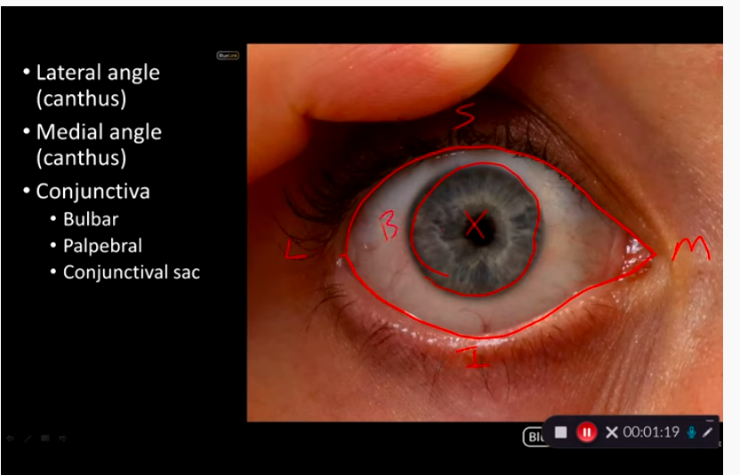
FIGURE-1
SOURCE OF YOU- https://www.youtube.com/watch?v=Ru9NTtMLe0k
Lacrimal anatomy (where tears go)
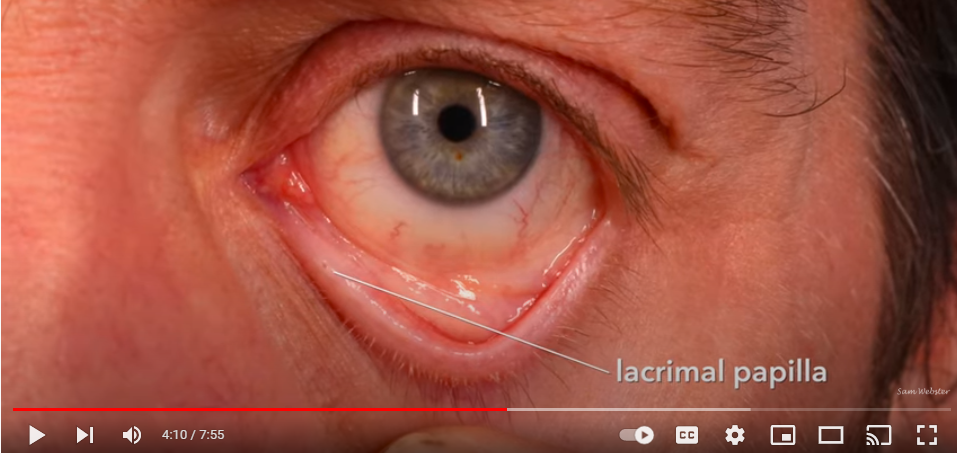
FIGURE 2
SOURCE OF THE FIGURE YOU TUBE-
SOURCE OF YOUTUBE LINK- https://www.youtube.com/watch?v=0HF5b-THh-A
Lacrimal anatomy (where tears go)
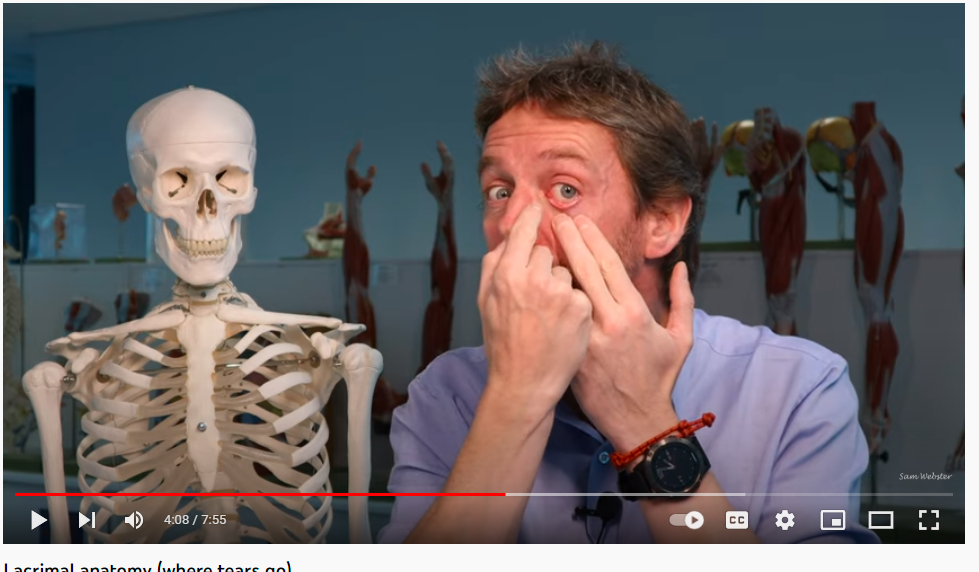
FIGURE-2
SOURCE OF YOUTUBE LINK- https://www.youtube.com/watch?v=0HF5b-THh-A
NASOLACRIMAL APPARATUS TEXT
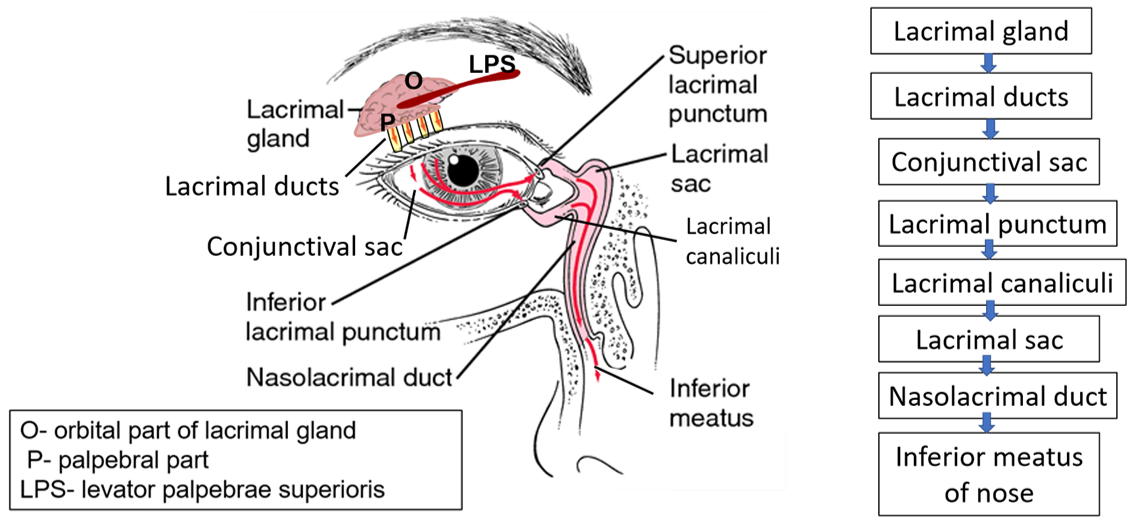
FIGURE-3
SOURCE OF FIGURE TEXT LINK- https://anatomyqa.com/lacrmal-apparatus-gland/
ANATOMY OF NASO LACRIMAL APPARATUS
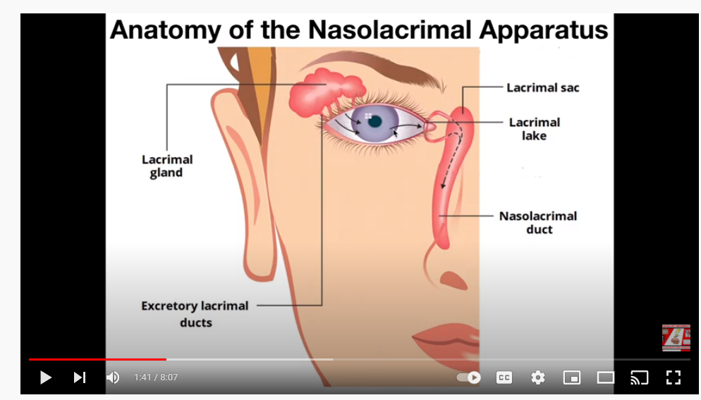
Figure-4
Source of you tube link- https://www.youtube.com/watch?v=TZg1L9cxdAg
MEATUS OF NOSE PHOTO
SCIENCE PHOTO LIBRARY
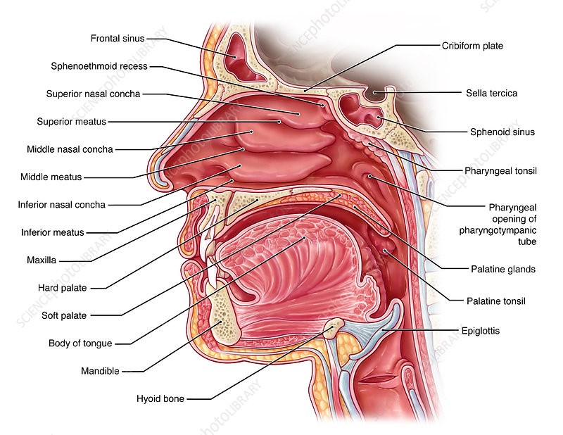
FIGURE-5
SOURCE OF PHOTO LINK- SCIENCE PHOTO LIBRARY
https://www.sciencephoto.com/media/861210/view
OPENING OF NASOLACRIMAL DUC ON INFERIOR MEATUS OF NOSE
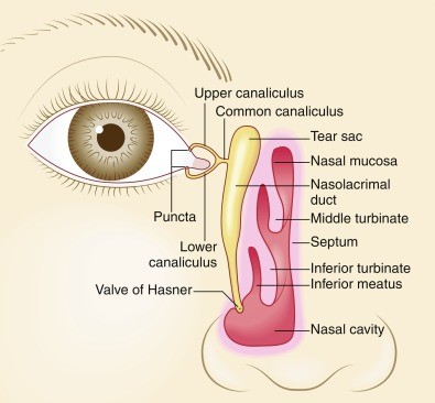
FIGURE -6
SOURCE OF PHOTO LINK– https://www.sciencephoto.com/media/861210/view
Special Senses | Eye Anatomy
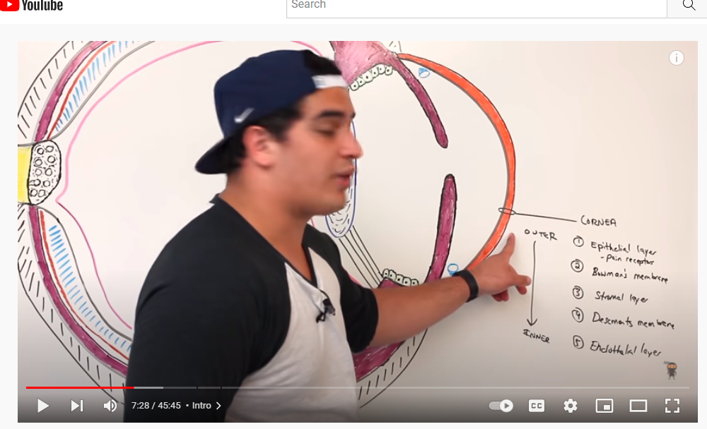
FIGURE-7
SOURCE OF YOU TUBE LINK- https://www.youtube.com/watch?v=Hclc7lL_Oyw
Special Senses | The Phototransduction Cascade YOU TUBE
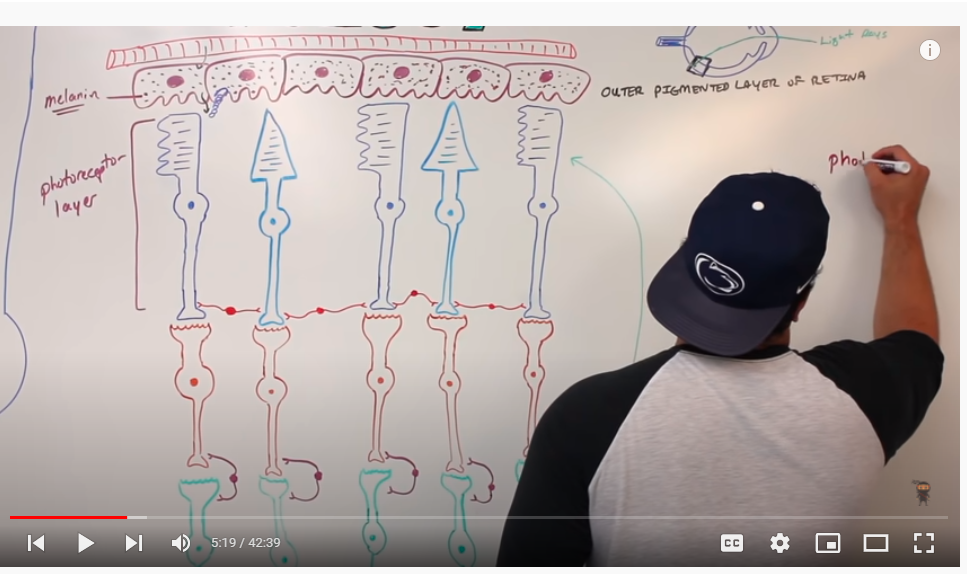
FIGURE-8
SOURCE OF YOU TUBE LINK– https://www.youtube.com/watch?v=GKaFjw8N8zQ&list=LLwbBmvre5US6kA7XCASuXyQ
Neurology | Optic Nerve | Cranial Nerve II: Visual Pathway and Lesions
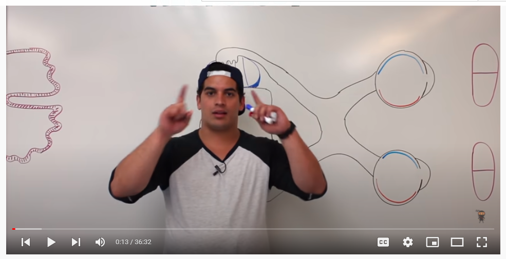
Figure-9
Source of youtube- https://www.youtube.com/watch?v=FnCiLD4gARI&list=LLwbBmvre5US6kA7XCASuXyQ
The Electromagnetic and Visible Spectra TEXT
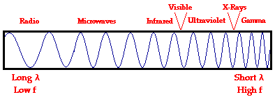
FIGURE 10
SOURCE OF TEXT LINK– https://www.physicsclassroom.com/class/light/u12l2a.cfm
DATE-5/14/2022
BLOG LINK
CHAPTER 31, EYE, HOW GLAUCOMA DEVELOPS.
[ ইংরেজীতে প্রকাশিত করা হল কিছু কিছু বাংলাদেশী বংশোদ্ভূত বিদেশে অবস্থানরত ছাত্র পাঠকেরা যারা বাংলা ভাষা পড়তে পারেনা, তাদের অনুরোধে, দুখিত ]
Dear viewers,
It is written as a primary knowledge for the students who are interested in studying medical sciences.
It may be somewhat useful for them. Today we are going to discuss about the way how the silent killer Glaucoma develops.
Aqueous Humour Eye Circulation Flow Animation: Open-Angle vs Closed-Angle Glaucoma

FIGURE-1
SOURCE OF YOU TUBE LINK- https://www.youtube.com/watch?v=O4lnSwZ8vFc
Development of Glaucoma Animation, Open Angle vs Angle Closure Glaucoma.
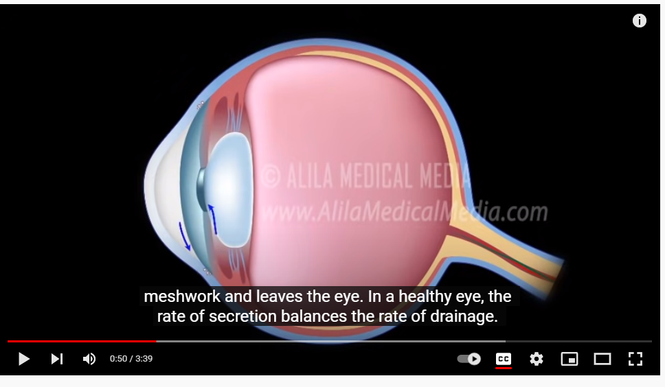
FIGURE-2
SOURCE OF YOU TUBE LINK- https://www.youtube.com/watch?v=TgjdPgSxeYg
Aqueous Humor Flow and Function TEXT
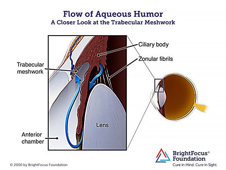
FIGURE-3
SOURCE OF THE TEXT- https://www.brightfocus.org/glaucoma/infographic/aqueous-humor-flow-and-function
Glaucoma TEXT
What is it?
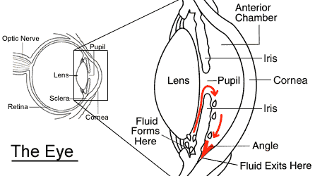
FIGURE-4
SOURCE OF THE TEXT- https://faculty.washington.edu/chudler/glaucoma.html
View a Video of the Eye’s Drainage System
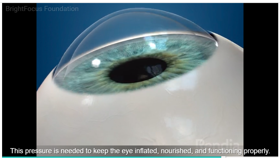
FIGURE 5
SOURCE OF YOU TUBE LINK- https://www.brightfocus.org/glaucoma/article/glaucoma-and-importance-eyes-drainage-system
Eyeball with Retina
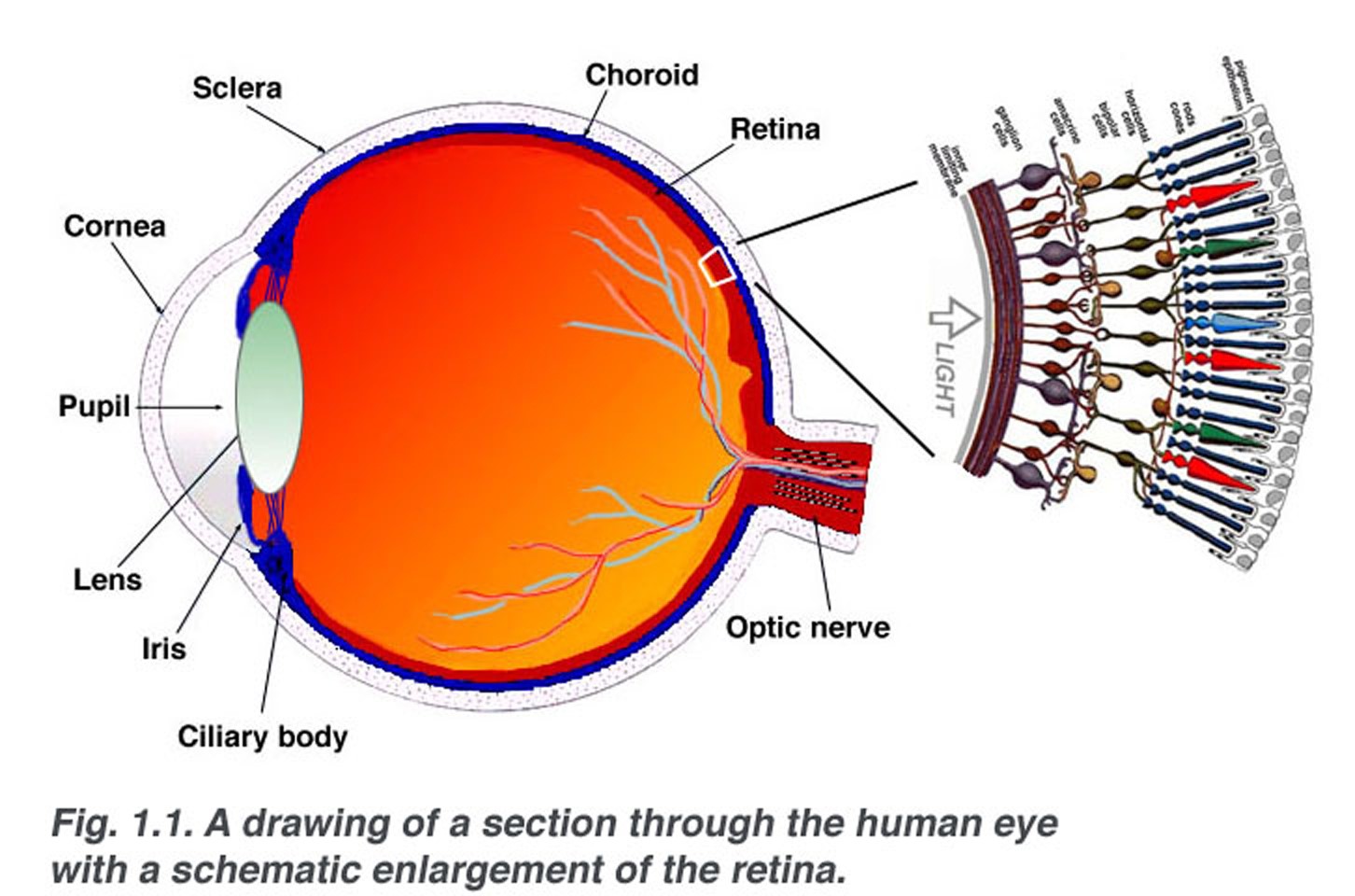
Figure 6
Source of the link- https://webvision.med.utah.edu/wp-content/uploads/2018/05/sagschem.jpg
DATE-
BLOG LINK –
CHAPTER 30, THE WAY WE CAN SEE. WHAT ARE THE PARTS OF OUR VISUAL SYSTEM? WHAT ARE CONVEX AND CONCAVE LENSES? HOW THEY REFRACT LIGHT?
[ ইংরেজীতে প্রকাশিত করা হল কিছু কিছু বাংলাদেশী বংশোদ্ভূত বিদেশে অবস্থানরত ছাত্র পাঠকেরা যারা বাংলা ভাষা পড়তে পারেনা, তাদের অনুরোধে, দুখিত ]
Dear viewers,
It is written as a primary knowledge for the students who are interested in studying medical sciences.
It may be somewhat useful for them. Today we are going to discuss about the way we can see.
ORBITAL CAVITY TEXT
SOURCE OF LTEXT LINK- https://en.wikipedia.org/wiki/Inferior_orbital_fissure
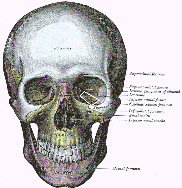
FIGURE-1
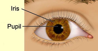
figure 2
Figure source-http://thebrain.mcgill.ca/flash/d/d_02/d_02_cr/d_02_cr_vis/d_02_cr_vis.html
1:38 / 3:32
Human Eye Structure and Functions || Human eye video for kids

FIGURE-3
SOURCE YOU TUBE LINK- https://www.youtube.com/watch?v=efgjoKNnwdw
How the Eye Works
Animation – How Do We See Video – Nearsighted & Farsighted Human
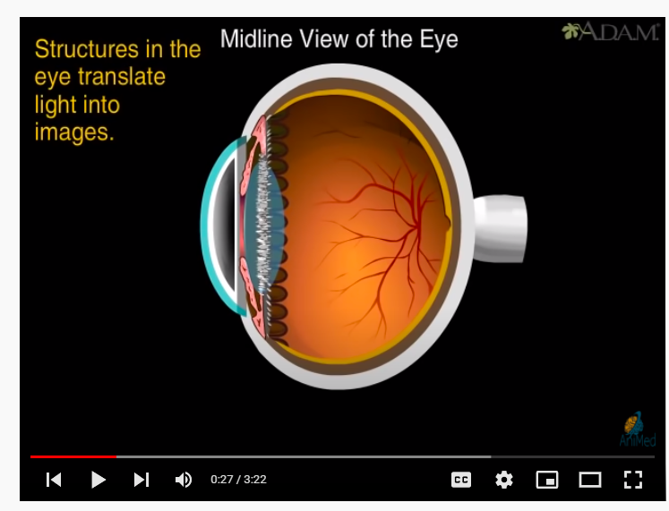
FIGURE-4
SOURCE OF YOUTUBE LINK- https://www.youtube.com/watch?v=YcedXDN6a88
Concave Convex lenses
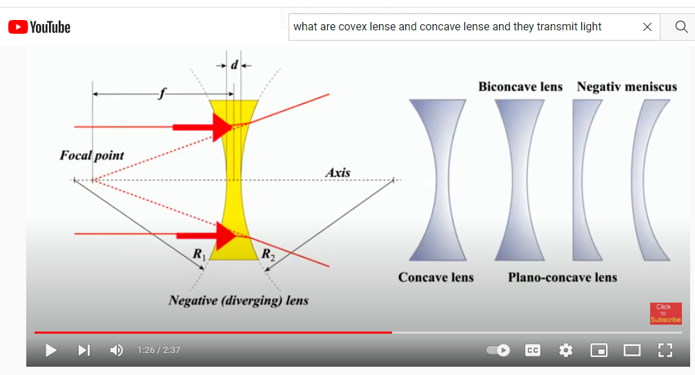
Figure 5
Source of you tube link- https://www.youtube.com/watch?v=oBLJ62uXRSc
EYE WORKS LIKE CAMERA
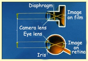
FIGURE 6
SOURCE OF LINK CHAPTER 21 FROM BLOG
Refraction of light through concave and convex lenses video
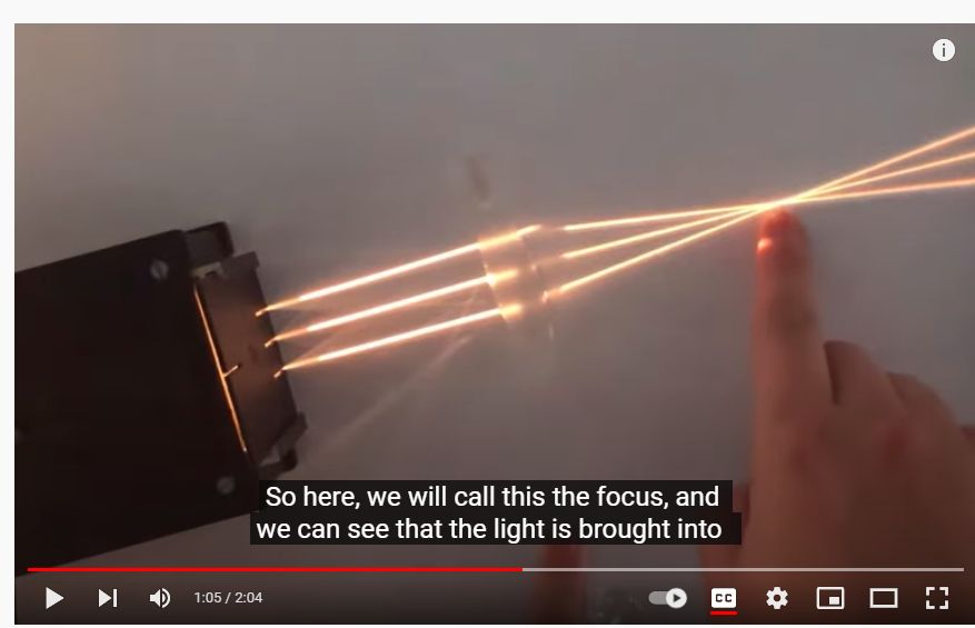
FIGURE-7
SOURCE OF YOU TUBE LINK- https://www.youtube.com/watch?v=_CT-wAIfOYQ
REFRACTION OF LIGHT THROUGH CONVEX LENCE
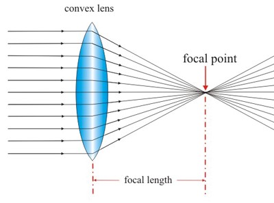
FIGURE-8
SOURCE OF FIGURE
CHAPTER 22- https://dnaandthemystryofhumanbody.wordpress.com/wp-admin/post.php?post=267&action=edit
DATE- 5/01/2022
BLOG LINK
