
CHAPTER 16, WHAT IS THE SPLEEN, WHAT IS IT’S FUNCTION?
[ ইংরেজীতে প্রকাশিত করা হল কিছু কিছু বাংলাদেশী বংশোদ্ভূত বিদেশে অবস্থানরত ছাত্র পাঠকেরা যারা বাংলা ভাষা পড়তে পারেনা, তাদের অনুরোধে, দুখিত ]
Dear viewers,
It is written as a primary knowledge for the students who are interested in studying medical sciences.
It may be somewhat useful for them.
Today we are discussing about the organ Spleen, and it’s functions.
Please open the You Tube links and figure links to understand clearly.

FIGURE- 1
Source of figure- https://radiopaedia.org/articles/abdominal-surface-anatomy-2
BLOOD SUPPLY OF SPLEEN AND DIGESTIVE SYSTEM AND SPLEEN

Figure-2
Source of the figure- ?
FUNCTION AND STRUCTURE OF SPLEEN

Figure-3
Source of figure text- https://www.news-medical.net/health/Role-of-the-Spleen-in-Drug-Metabolism.aspx
YOU TUBE –FUNCTIONS & STRUCTURE OF SPLEEN
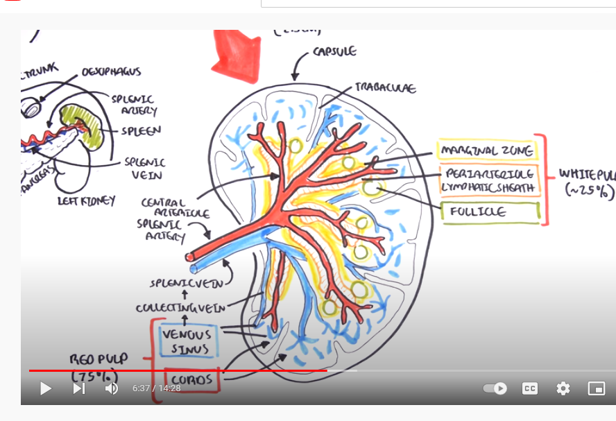
FIGURE- 4
SOURCE OF YOU TUBE LINK- https://www.youtube.com/watch?v=RezL2xWFCe8&t=5s
FUNCTION OF SPLEEN- YOU TUBE

FIGURE-5
SOURCE OF FIGURE- https://www.youtube.com/watch?v=ah74jT00jBA
STRUCTURE OF SPLEEN
WHAT IS THE FUNCTION OF THE SPLEEN?
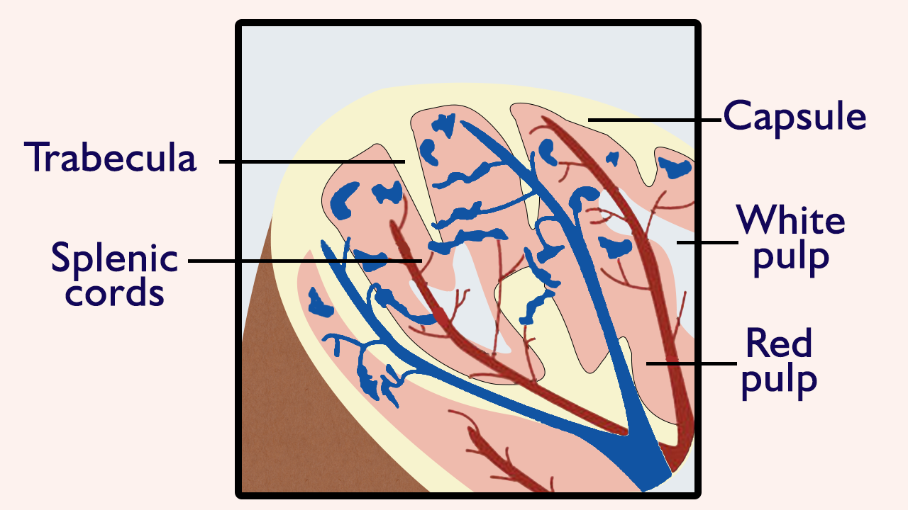
FIGURE- 6
SOURCE OF FIGURE- https://microbenotes.com/spleen-structure-and-functions/
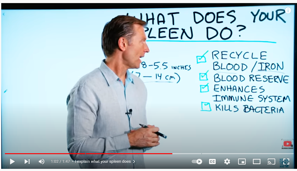
FIGURE-7
LINK OF THE YOU TUBE- https://www.youtube.com/watch?v=kiXbC0L-e4g
DATE- 1/30/2022
BLOG LINK-
CHAPTER 15, WHAT IS THE PANCREAS, WHAT IS ITS FUNCTION? Date -1/29/2022
[ ইংরেজীতে প্রকাশিত করা হল কিছু কিছু বাংলাদেশী বংশোদ্ভূত বিদেশে অবস্থানরত ছাত্র পাঠকেরা যারা বাংলা ভাষা পড়তে পারেনা, তাদের অনুরোধে, দুখিত ]
Dear viewers,
It is written as a primary knowledge for the students who are interested in studying medical sciences.
It may be somewhat useful for them.
Today we are discussing about the organ Pancreas, and it’s functions.
Please open the You Tube links and figure links to understand clearly.
9 QUADRATE OF ABDOMEN ON SUFACE

FIGURE- 1
Source of figure- https://radiopaedia.org/articles/abdominal-surface-anatomy-2
BLOOD SUPPLY OF SPLEEN AND DIGESTIVE SYSTEM AND SPLEEN
BLOOD SUPPLY OF DIGESTIVE ORGANS
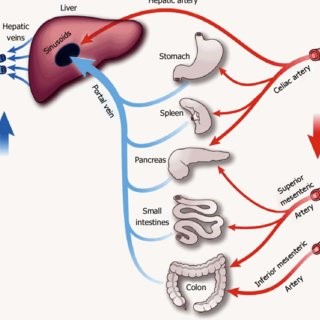
FIGURE-2
SOURCE OF FIGURE- ?
YOU TUBE –FUNCTION & STRUCTURE OF PANCREAS
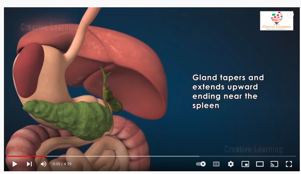
FIGURE-3
SOURCE OF YOU TUBE LINK- https://www.youtube.com/watch?v=bqpHnOcWvdM
STRUCTURE AND FUNCTION OF PANCREAS IN TEXT
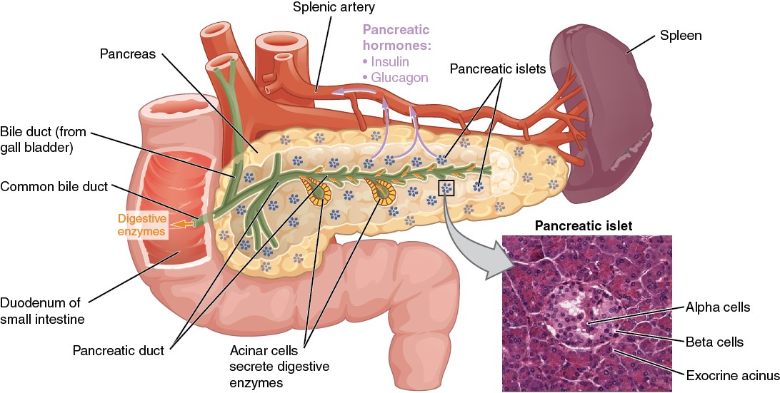
FOGURE-4
SOURCE OF FIGURE AND TEXT – https://open.oregonstate.education/aandp/chapter/17-9-the-pancreas/
DATE- 1/29/2022
BLOG LINK-
CHAPTER 14, WHAT ARE LIVER, ITS PORTAL VENOUS SYSTEM, PORTAL HYPERTENSION AND GALL BLADDER?
Date -1/22/2022
[ ইংরেজীতে প্রকাশিত করা হল কিছু কিছু বাংলাদেশী বংশোদ্ভূত বিদেশে অবস্থানরত ছাত্র পাঠকেরা যারা বাংলা ভাষা পড়তে পারেনা, তাদের অনুরোধে, দুখিত ]
Dear viewers,
It is written as a primary knowledge for the students who are interested in studying medical sciences.
It may be somewhat useful for them.
Today we are discussing about the organ Liver, gall bladder, and their functions.
Please open the You Tube links and figure links to understand clearly.
What is the gallbladder and what does it do?
9 QUADRANT OF ABDOMEN-Abdominal surface anatomy,
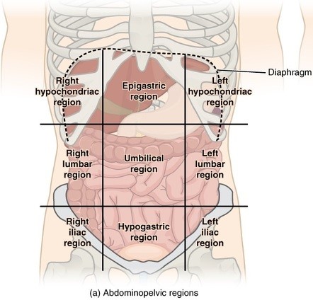
FIGURE- 1
Source of figure- https://radiopaedia.org/articles/abdominal-surface-anatomy-2
BLOOD CIRCULATION OF BODY
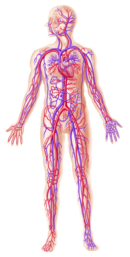
FIGURE- 2
SOURCE OF FIGURE-
BLOOD CIRCULATION OF BODY-
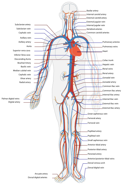
Figure-3
ABDOMINAL BLOOD CIRCULATION\

FIGURE- 4
SOURCE OF THE FIGURE-
https://slidetodoc.com/large-blood-vessels-of-the-gut-the-coeliac/
VENOUS DRAINAGE OF LIVER, STOMACH, SPLEEN, PANCREAS, SMALL INTESTINE, AND COLON

Figure-5
Sorce of figure- ?
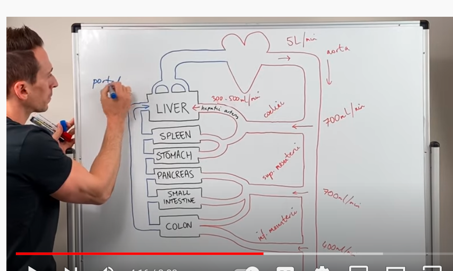
FIGURE-6
SOURCE OF THE YOU TUBE- Hepatic Circulation (Liver Blood Supply) – YouTube
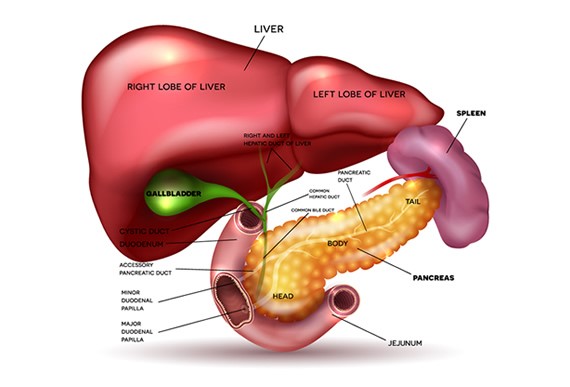
Figure 7
SOURCE OF FIGURE-
(go through it for good picture and detail ) http://www.hpblondon.com/gallstones/
ORDINARY CAPILLARY BED OF THE BODY
SOURCE OF FIGURE- BELOW
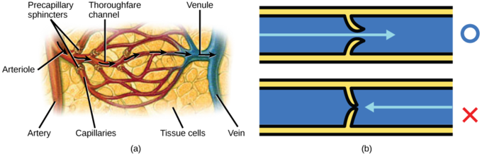
FIGURE- 8
SOURCE OF THE FIGURE- https://courses.lumenlearning.com/boundless-biology/chapter/blood-flow-and-blood-pressure-regulation/
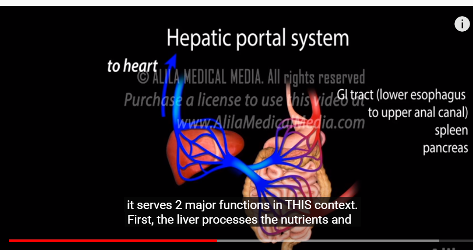
FIGURE-9
YOU TUBE LINK- https://www.youtube.com/watch?v=mMKdUgNdhBs
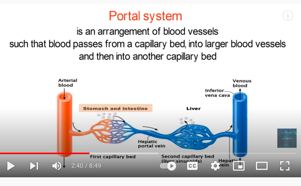
Figure-10
YOU TUBE LINK- Hepatic Portal System! – YouTube (VERY IMPORTANT)
PORTAL VENOUS SYSTEM
FIGURE-11
YOU TUBE LINK-
Hepatic portal vein (anatomy) – YouTube
POTAL VENOUS SYSTEM
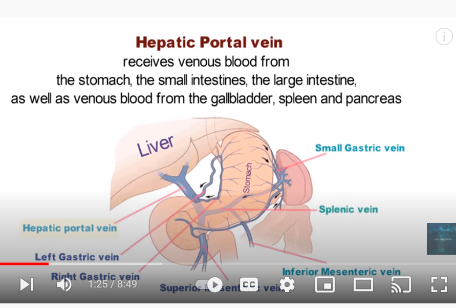
FIGURE-12
PORTAL VENOUS SYSTEM
PORTAL VENOUS SYSTEM
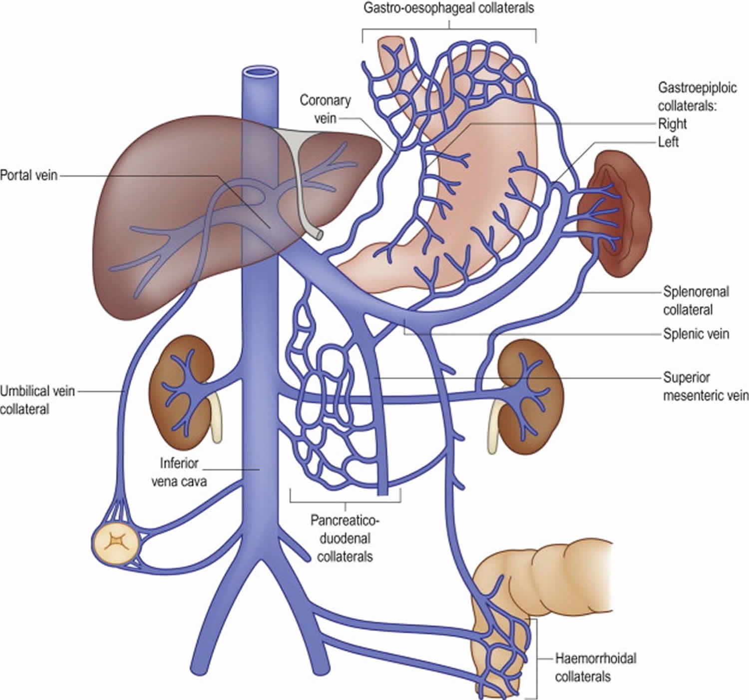
FIGURE-13
SOURCE OF LINK – https://www.verywellhealth.com/portal-vein-anatomy-4689616 (VERY IMPORTANT)
HEPATIC PORTAL DRAINAGE-
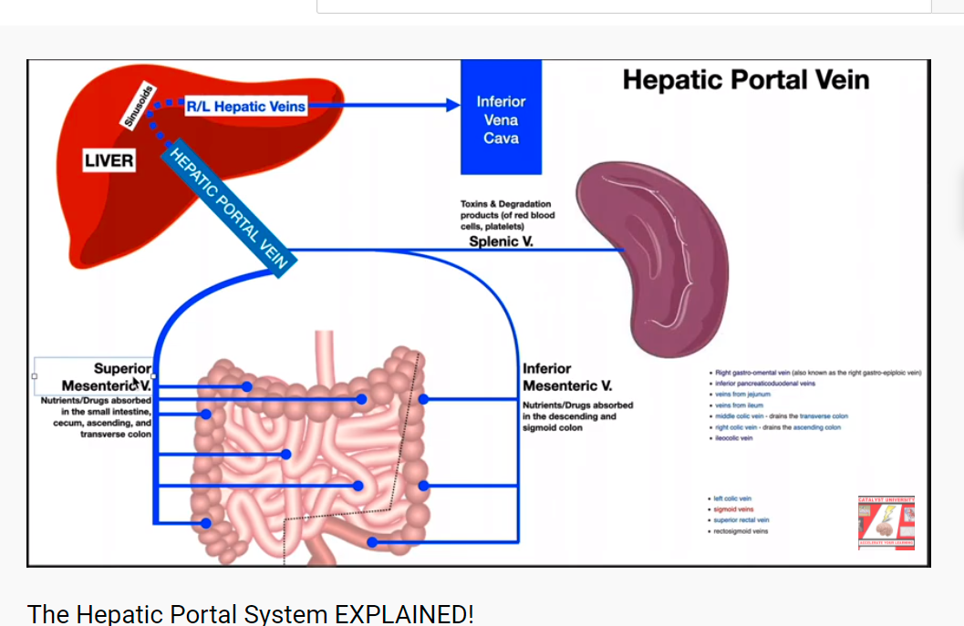
FIGURE-14
YOU TUBE LINK- The Hepatic Portal System EXPLAINED! – YouTube
BLOOD DRAINAGE OF GASRO INTESTINAL (GI) TRACT TO LIVER THROUGH PORTAL VEIN.
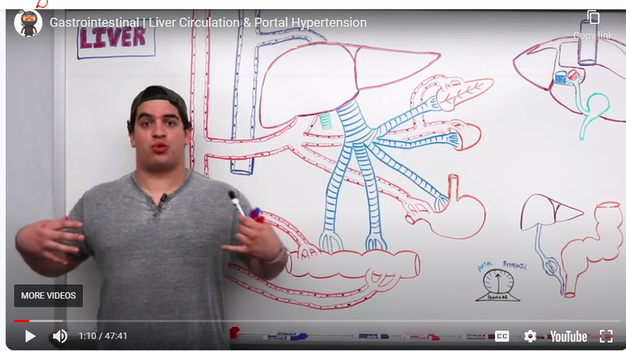
FIGURE-15
You tube link– https://www.ninjanerd.org/lecture/liver-circulation-portal-hypertension
The Portal Circulation
The liver is unusual in that it has a double blood supply; the right and left hepatic arteries carry oxygenated blood to the liver, and the portal vein carries venous blood from the GI tract to the liver.
The venous blood from the GI tract drains into the superior and inferior mesenteric veins; these two vessels are then joined by the splenic vein just posterior to the neck of the pancreas to form the portal vein. This then splits to form the right and left branches, each supplying about half of the liver.
On entering the liver, the blood drains into the hepatic sinusoids, where it is screened by specialised macrophages (Kupffer cells) to remove any pathogens that manage to get past the GI defences. The plasma is filtered through the endothelial lining of the sinusoids and bathes the hepatocytes; these cells contain vast numbers of enzymes capable of braking down and metabolising most of what has been absorbed.
The portal venous blood contains all of the products of digestion absorbed from the GI tract, so all useful and non-useful products are processed in the liver before being either released back into the hepatic veins which join the inferior vena cava just inferior to the diaphragm, or stored in the liver for later use.
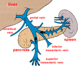
FIGURE-16
SOURCE OF THE FIGURE- https://www.le.ac.uk/pa/teach/va/anatomy/case5/5_3.html
FUNCTIONS OF LIVER, HOW TO CLEANSE YOUR LIVER , KIDNEY, MECHANISM OF URINE FORMATION.
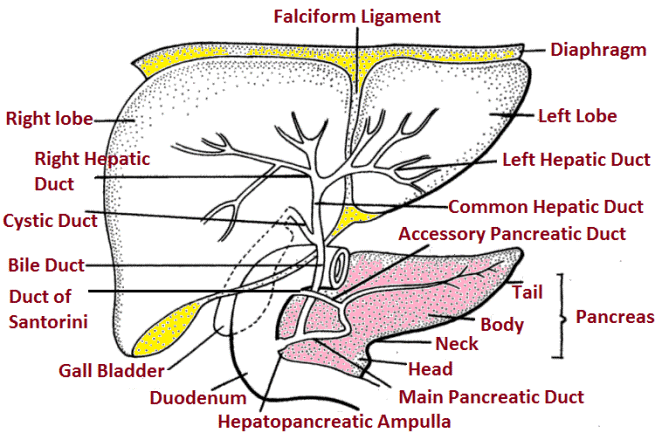
FIGURE-17
SOURCE OF FIGURE- (SEE STRUCTURE AND FUNCTIONS OF LIVER)
FUNCTIONS OF LIVER:
STRUCTURE OF LIVER
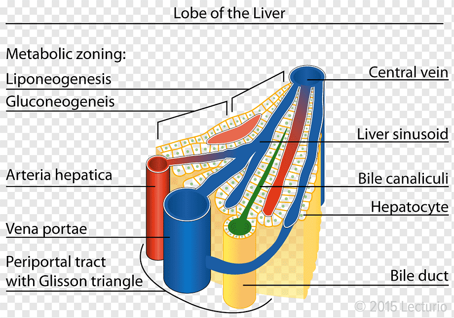
FIGURE-18
SOURCE OF FIGURE- https://www.pngwing.com/en/free-png-ieoya
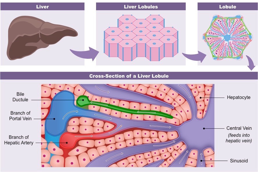
FIGURE-19
Hepatic Lobules- SINUSOIDS OF LIVER
REFERENCE & SOURCE OF FIGURE- https://ib.bioninja.com.au/options/option-d-human-physiology/d3-functions-of-the-liver/liver-structure.html
Venous drainage of liver, stomach, small intestine, lage intestine, spleen and pancreas.
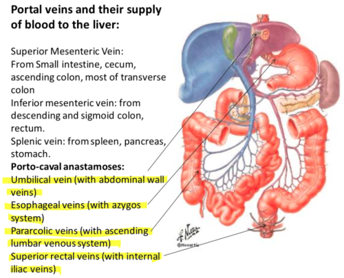
Figure-20
Surce of the figure- https://quizlet.com/130121255/veinous-drainage-of-the-abdomen-flash-cards/
VENOUS DRAINAGE OF LIVER, STOMACH, PANCREAS, SPLEEN, SMALL INTESTINE AND COLON.
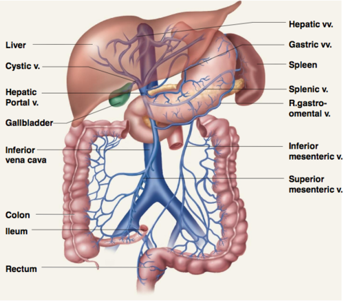
FIGURE-21
SOURCE OF THE FIGURE- https://quizlet.com/71372414/bio-202-lab-4-hepatic-portal-vein-flash-cards/
PORTAL HYPERTENTION, CIRRHOSIS LIVER, VARIECES , AND BLEEDING IN STOMACH OR INTESTINES. YOU TUBE.
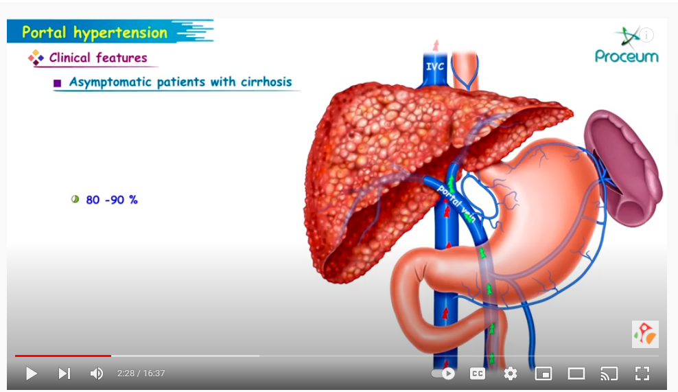
FIGURE-22
SOURCE OF YOU TUBE LINK– https://www.youtube.com/watch?v=cbq12NU3Uj0
PORTAL HYPERTENTION, CIRRHOSIS LIVER, VARIECES , AND BLEEDING IN STOMACH OR INTESTINES. YOU TUBE.
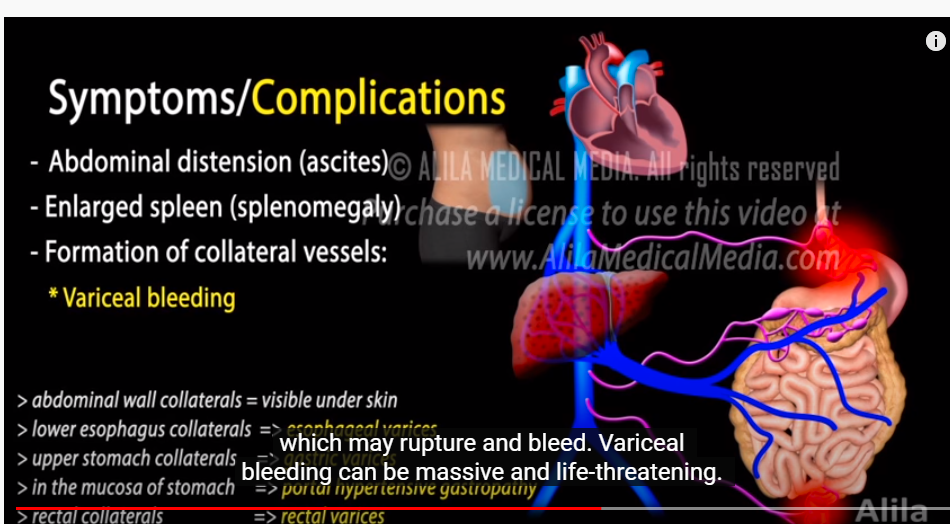
FIGURE-23
SOURCE OF FIGURE- https://www.youtube.com/watch?v=LkXQTDb8g2U
DATE- 1/22/2022
BLOG LINK-
CHAPTER 13, WHAT ARE THE PARTS OF DIGESTIVE SYSTEM? HOW DOES IT WORK?
CHAPTER 13, WHAT ARE THE PARTS OF DIGESTIVE SYSTEM? HOW DOES IT WORK?
[ ইংরেজীতে প্রকাশিত করা হল কিছু কিছু বাংলাদেশী বংশোদ্ভূত বিদেশে অবস্থানরত ছাত্র পাঠকেরা যারা বাংলা ভাষা পড়তে পারেনা, তাদের অনুরোধে, দুখিত ]
Dear viewers,
It is written as a primary knowledge for the students who are interested in studying medical sciences.
It may be somewhat useful for them.
Today we are discussing about The Digestive System, what its parts are and how it works.
Please open the You Tube links and figure links to understand clearly.
PAROTID (SALIVERY) GLAND
FIGURE-1
YOU TUBE LINK- https://www.youtube.com/watch?v=HB6bN-rs2NU

FIGURE-2
Source of figure- https://www.healthdirect.gov.au/digestive-system
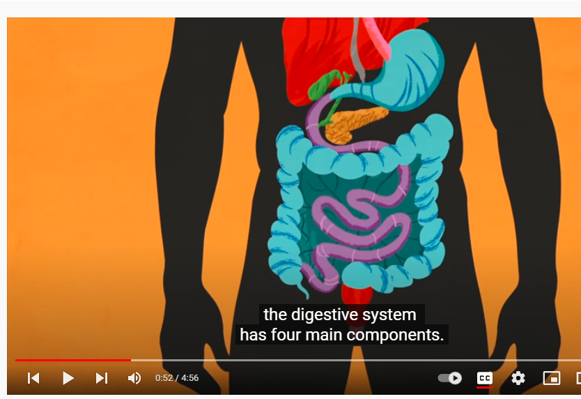
Figure-3
You tube link-
https://www.youtube.com/watch?v=Og5xAdC8EUI&t=17s
PERITONIUM MESENTRY
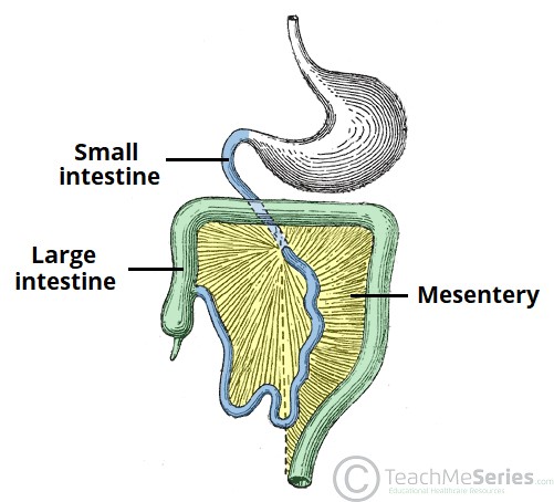
FIGURE- 4
FIGURE- SOURCE OF THE FIGURE https://teachmeanatomy.info/abdomen/viscera/mesentery/
PERITONIUM GREATER AND LESSER OMENTUM
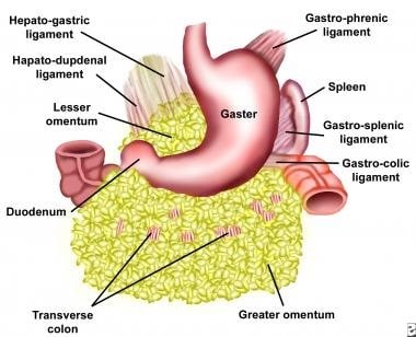
FIGURE-5
SOURCE OF THE FIGURE-https://emedicine.medscape.com/article/193622-overview
LAYERS OF DIGESTIVE TRACT-
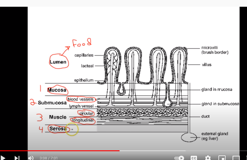
FIGURE-6
LINK TO YOU TUBE-
https://www.youtube.com/watch?v=VSPdLCwfMu8&t=150s
WALL OF THE DIGESTIVE TRACT
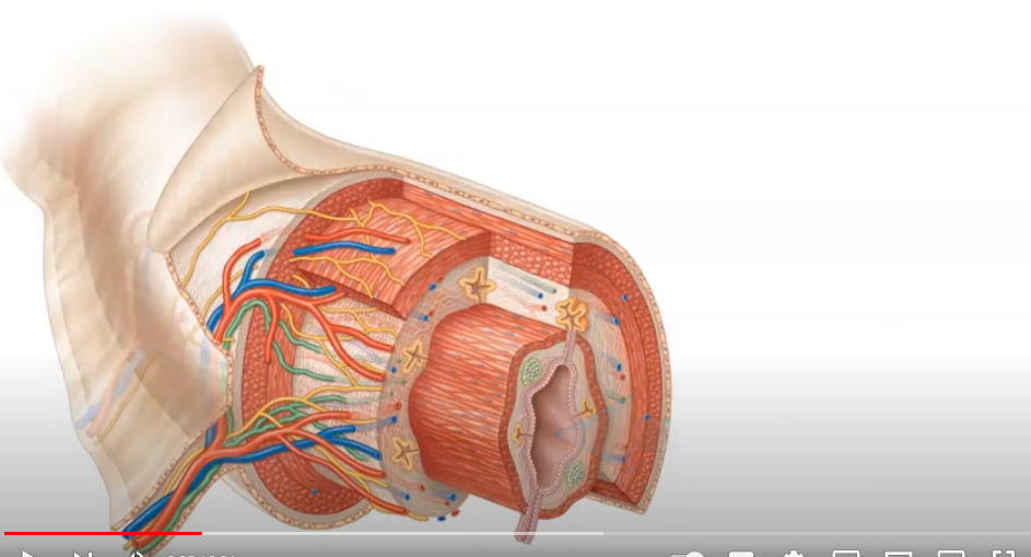
FIGURE-7
LINK TO YOU TUBE-
https://www.youtube.com/watch?v=7rFrnnrus7Y
HOW MANY TYPES OF MAIN FOOD GROOPS ARE?
Answer- Three main types-
- CARBOHYDRATES- as rice, bread, cereals, fruits etc.
- PROTENS- as meat, fish ,egg, milk etc.
- FATS- as butter, ghee, oils, egg yolk, milk etc.
HOW MUCH ENERGY IS GIVEN BY FOOD?
Answer-
1 gram of carbohydrate gives 4 calories
1 gram of protein gives 4 calories
1 gram of fat gives 9 calories
How many types of carbohydrates are structurally?
5 types-
- POLYSACHARIDE- It contains many molecules of monosaccharide-
- Oligosaccharide- it contains 3-6 monosaccharide-
- Disaccharide – it contains 2 molecules of monosaccharaides-
- Nucleotide- it is a component of nucleic acid or DNA
- It is a single component of carbohydrate- they are three types- 1. Glucose. 2. Fructose. 3. Galactose.
So the end product of polysaccharides are monosaccharide.
Chemical structure of polysaccharide disaccharide, and monosaccharide-

Figure-8
Source of figure
https://www.scienceofcooking.com/chemical_physical_properties_polysaccharides.htm
–
WHAT ENZYMES BREAK DOWN CARBOHYDRATE INTO MONOSACCHARAIDES (GLUCOSE)?
Answers-
- SALIVARY AMYLASE- secreted in mouth by chewing.
- PANCREATIC AMYLASE secreted by pancreas.
- MALTASE, SUCRASE AND LACTASE- secreted by intestinal glands.
Source for links– https://healthyeating.sfgate.com/steps-digestion-carbohydrates-4053.html
PROTEIN
Proteins are composed of AMINO ACIDS or Proteins are formed by joining of Amino acid through peptide bonds.
So the end products of proteins are Amino acids.
Chemical Structure of an amino acid-
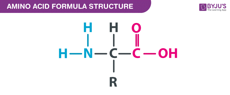
Figure-9
Source of figure- https://byjus.com/amino-acid-formula/
Chemical structure of a protein-
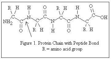
Figure-10
Source of figure-
http://milkfacts.info/Milk%20Composition/Protein.htm
WHAT ENZYMES BREAK DOWN PROTEINS?
Answer-
- Protease and hydrochloric acid secreted from stomach.
- From intestine- Trypsine, Chymotrypsine, carboxy peptidase
Source for link- https://www.healthline.com/health/protein-digestion#enzymes
chemical structure of a fatty acid-
Chemical structure of a fat
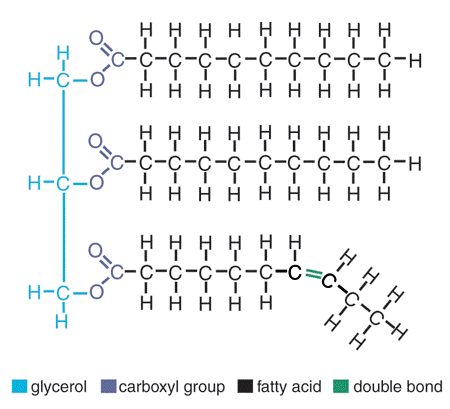
Figure-11
Source of figure- https://www.visionlearning.com/en/library/Biology/2/Lipids/207
Figure-
Source of figure- https://courses.lumenlearning.com/pierce-nutritionmaster/chapter/essential-fatty-acids/
WHAT ARE THE ENZYMES BREAKDOWN FATS?
Answer-
- Lipase – secreted from stomach and Pancreas.
- Bile- secreted from liver
Source of link-
https://therahealth.com.au/enzymes-for-fat-digestion/
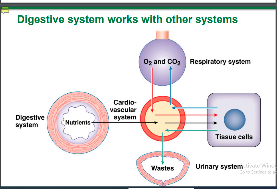
Figure- 12
Source of figure- https://www.palmbeachstate.edu/slc/Documents/AandPch22LecturePearson.pdf
DATE- 01/15/2022
BLOG LINK-
CHAPTER 12, RESPIRATORY SYSTEM, HOW IT WORKS AND WHAT ARE ITS PARTS? WHAT IS PLEURAL SAC?WHAT IS THE RELATION OF THE UPPER PART OF DIGESTIVE SYSTEM?
CHAPTER 12
RESPIRATORY SYSTEM, HOW IT WORKS AND WHAT ARE ITS PARTS? WHAT IS PLEURAL SAC?WHAT IS THE RELATION OF THE UPPER PART OF DIGESTIVE SYSTEM?
[ ইংরেজীতে প্রকাশিত করা হল কিছু কিছু বাংলাদেশী বংশোদ্ভূত বিদেশে অবস্থানরত ছাত্র পাঠকেরা যারা বাংলা ভাষা পড়তে পারেনা, তাদের অনুরোধে, দুখিত ]
Dear viewers,
It is written as a primary knowledge for the students who are interested to study medical sciences.
It may be somewhat useful for them.
Today we are discussing about respiratory system, what its parts are and how it works,
What its blood and nerve supply are.
Please open the You Tube links to understand clearly.
You tube link
https://www.youtube.com/watch?v=kacMYexDgHg
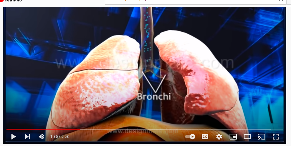
Figure-1
PLEURAL SAC
You Tube Link- https://www.youtube.com/watch?v=dHxhLRCA4v8
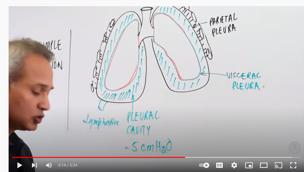
Figure- 2
PLEURAL SAC
YOU TUBE LINK
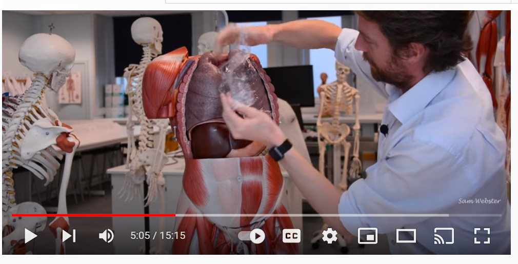
FIGURE- 3
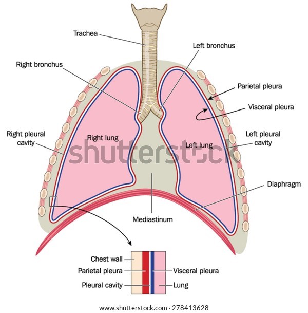
Figure- 4
Source of figure- https://www.shutterstock.com/image-vector/lungs-trachea-bronchi-mediastinum-detail-chest-278413628
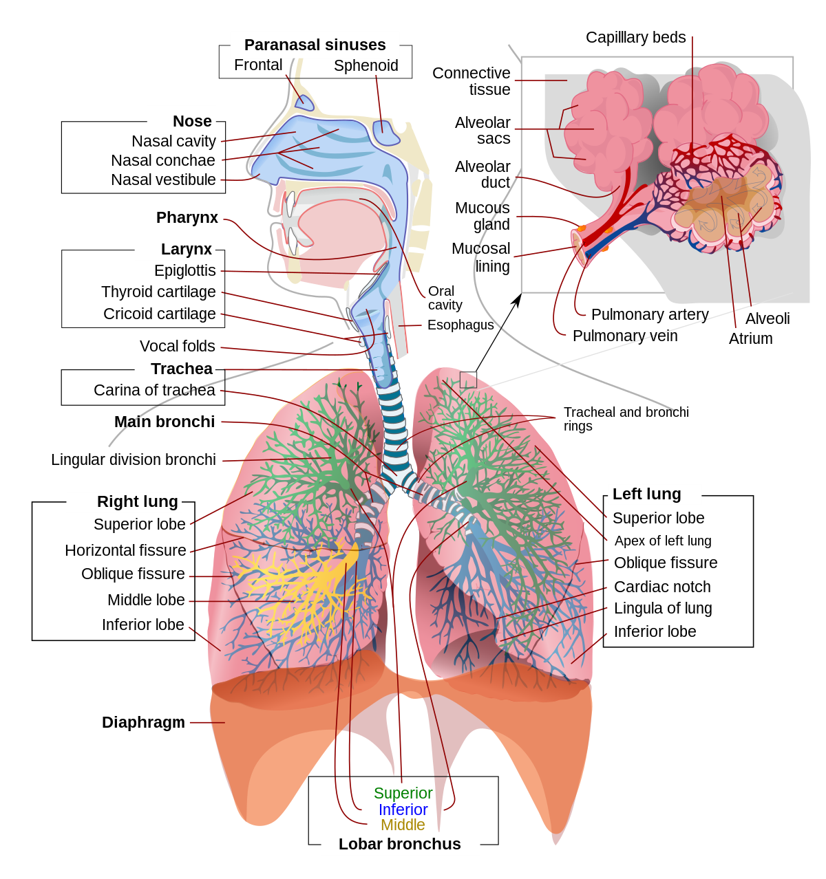
Figure-5
Source of figure– https://en.wikipedia.org/wiki/Respiratory_system
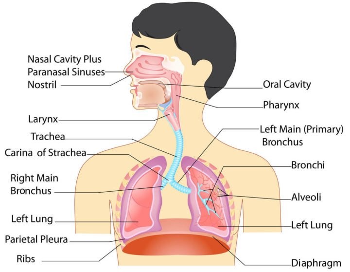
Figure- 6
Source of figure- https://adrenalfatiguesolution.com/anatomy-of-the-respiratory-system/
LARYNX AND PHARYNX RELATION
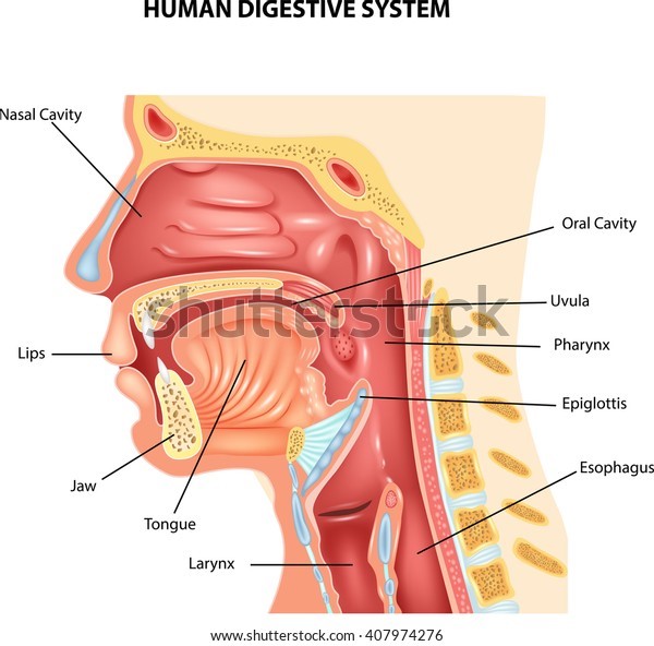
FIGURE- 7
SOURCE OF FIGURE- (HUMAN DIGESTIVE SYSTEM) – https://www.shutterstock.com/image-vector/illustration-human-digestive-system-407974276
DIGESTIVE SYSTEM RELATION WITH RESPIRATORY SYSTEM
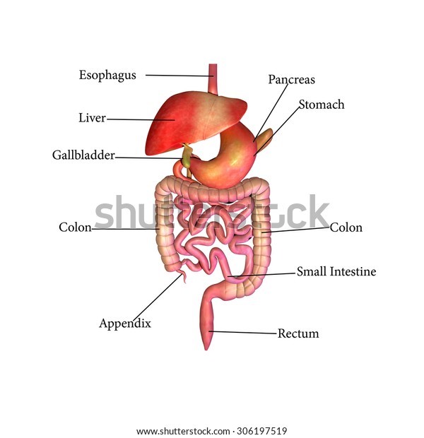
FIGURE-8
SOURCE OF FIGURE- Human Digestive System Stock Illustration 306197519 (shutterstock.com)
BRONCHIAL TREE
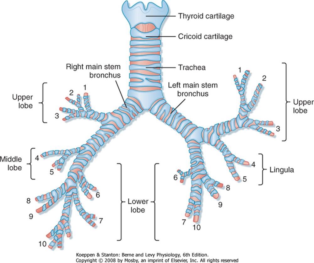
FIGURE-9
SOURCE OF FIGURE- https://doctorlib.info/physiology/physiology/20.html
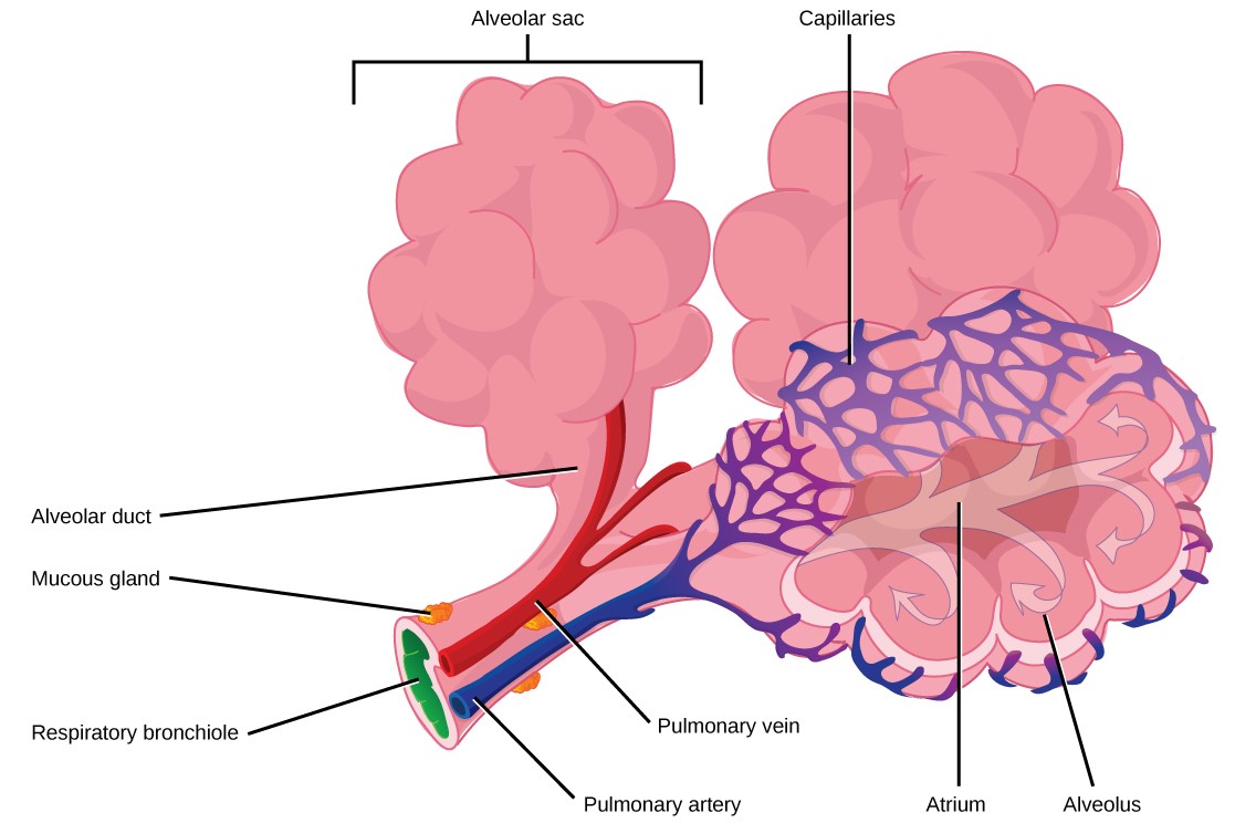
Figure-10
Source of figure- https://ecampusontario.pressbooks.pub/conceptsbiocdnremediate/chapter/11-3-circulatory-and-respiratory-systems/
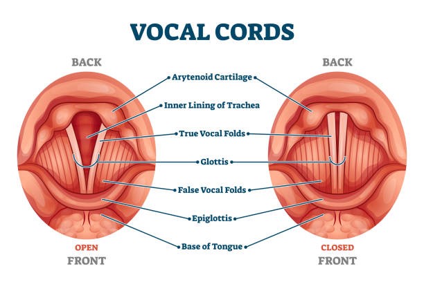 1
1
Source of figure- https://www.istockphoto.com/illustrations/vocal-cords
Date-01/08/2022
Blog link-
