
CHAPTER 8, ANATOMY OF HUMAN CIRCULATORY SYSTEM
[ ইংরেজীতে প্রকাশিত করা হল কিছু কিছু বাংলাদেশী বংশোদ্ভূত বিদেশে অবস্থানরত ছাত্র পাঠকেরা যারা বাংলা ভাষা পড়তে পারেনা, তাদের অনুরোধে, দুখিত ]
Dear viewers,
This is written as a primary knowledge for the students who are interested to study medical science.
It may be somewhat useful for them.
Please open the web links to understand clearly.
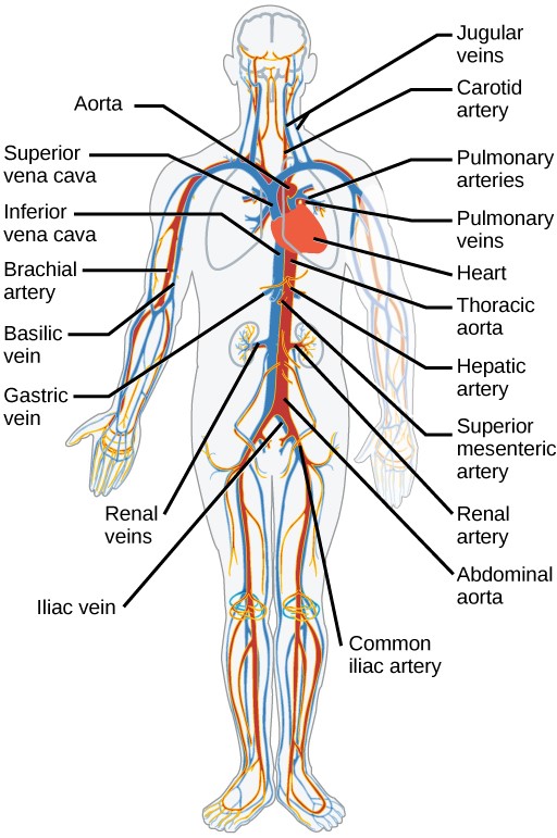
Figure-1
Source of figure- https://www.khanacademy.org/science/in-in-class-10-biology/in-in-life-processes/in-in-transportation-in-human-beings/a/hs-the-circulatory-system-review
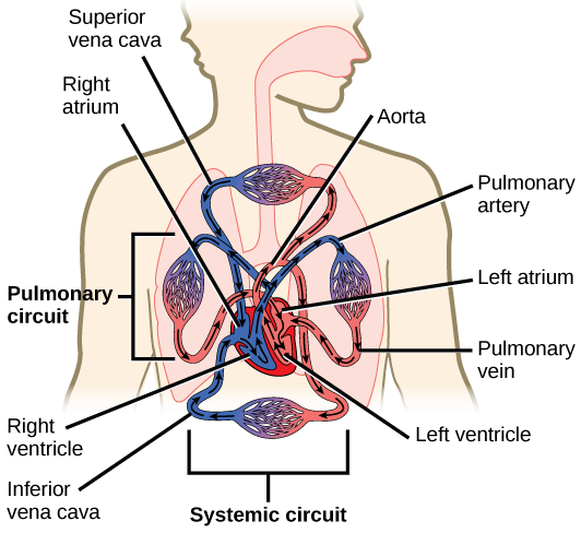
Figure -2
Source of figure- https://www.khanacademy.org/science/in-in-class-10-biology/in-in-life-processes/in-in-transportation-in-human-beings/a/hs-the-circulatory-system-review
Figure-3
Date-11/28/2021
Blog post-
web links-
CHAPTER 7, TETRALOGY OF FALLOT OR FALLOT’S TETRALOGY”
[ ইংরেজীতে প্রকাশিত করা হল কিছু কিছু বাংলাদেশী বংশোদ্ভূত বিদেশে অবস্থানরত ছাত্র পাঠকেরা যারা বাংলা ভাষা পড়তে পারেনা, তাদের অনুরোধে, দুখিত ]
Dear viewers,
This is written as a primary knowledge for the students who interested to study medical science.
It may be somewhat useful for them.
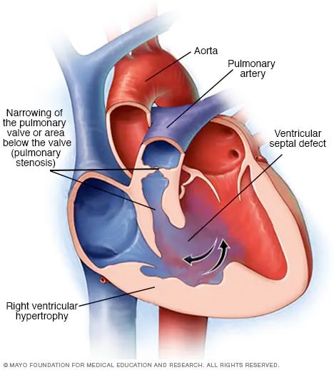
Today we are discussing about a congenital heart disease called ‘TETRALOGY OF FALLOT OR FALLOT’S TETRALOGY”
The following changes are found in the heart of the patient baby.
1.VENTRICULAR SEPTAL DEFFECT
2.OVERRIDING AORTA
3.PULMONARY STENOSIS
4.RIGHT VENTRICULAR HYPERTROPHY
Since there is a pore in the in the ventricular septum, the deoxygenated blood and oxygenated blood are mixed in together. So body cannot find fully oxygenated blood.
The patient suffers from cyanosis or blue color and complications
Please view the you tube link below to understand clearly with the animated video.
YOU TUBE LINK OF FALLOTS TETRELOGY
https://www.youtube.com/watch?v=8BJOUfycsxo
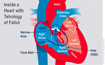
Figure 1
FIGURE=2
YOU TUBE LINK OF FALLOT’S TETRALOGY.
“Tetralogy of Fallot: Management Strategies,” by Peter Lang, MD, for OPENPediatrics – YouTube
Why a baby is attacked with this disease?
Unfortunately, if a baby receives some mutated genes like, NKX2-5[5]
ZFPM2[6] (1.4). etc.
You can see the location of gene, NKX2-5 below
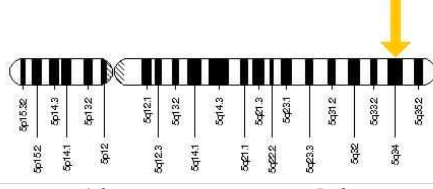
Figure-3
Chromosome 5
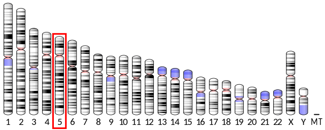
Figure-4
23 pairs of chromosome in human
Figure souce- https://en.wikipedia.org/wiki/Tetralogy_of_Fallot?fbclid=IwAR0b7Rb0qwoJTcLf98h5qovUnfmXQYhrljQ8r47_JBZIPGjZFX5UFupocW0
FIGURE
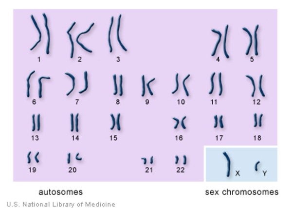
FIGURE-5
23 pairs of human chromosome (Kariotype)
FIGURE SOURCE-
https://medlineplus.gov/genetics/understanding/basics/howmanychromosomes/
Molecular Location on chromosome 5: base pairs 173,232,103 to 173,235,311
Mutation of this gene NKX2-5,can cause Fallots Tetralogy
This gene is located in chromosome 5 at q arm at 34 location.
More precisely, NKX2-5, is located from base pair 173,232,103 to 311 base pair 173,235, in chromosome
How many base pairs do have human chromosome?
Answer- about 3 billions.
YOU TUBE LINK-
A Face book link of a patient
https://www.facebook.com/abdul.chakladar.9/posts/842073719158127
DATE- 11/27/2017
BLOG LINK-
SOURCE OF FIGURE 1
https://kidshealth.org/Advocate/en/parents/tetralogy-of-fallot.html
SOURCE OF FIGURE 2
https://www.mayoclinic.org/diseases-conditions/tetralogy-of-fallot/symptoms-causes/syc-20353477
CHAPTER 6, CARDIAC CONDUCTION
CARDIAC CONDUCTION
DIFFERENCE BETWEEN AN ORDINARY ACTION POTENTIAL AND A MYOCARDIAL ACTION POTENTIAL AND RELATION BETWEEN MYOCARDIAL ACTION POTENTIAL CHARGE AND ECG OR EKG.
[ ইংরেজীতে প্রকাশিত করা হল কিছু কিছু বাংলাদেশী বংশোদ্ভূত বিদেশে অবস্থানরত ছাত্র পাঠকেরা যারা বাংলা ভাষা পড়তে পারেনা, তাদের অনুরোধে, দুখিত ]
Dear viewers,
This is written as a primary knowledge for the students who intend to study medical science.
It may be somewhat useful for them.
Today we are discussing how heart is conducted. We know about SA node located at upper part Rt. Atrium. It is a pace maker.it produces electric charge and transmits it to AV node. AV node transmits to Bundle of His and Bundle of His transmits through Purkenji Fibers to heart muscles.
The electric charge transmitted through Neuron with Action Potential in the body is not similar.
It occurs through different scales in different tissues.
Please look below a comparison between the depolarization, peak, repolarization resting stage and refractory period of an Action Potential of an ordinary neuron and that of a Cardiac muscle neuron.
SEE BELOW THE ACTION POTENTIAL CHARGE RELATION WITH ECG OR EKG.
Action potential of cardiac muscle and SA Node – Usmle step 1 CVS Physiology
U- tube link-
https://www.youtube.com/watch?v=-13IfTvjZRI&list=LLwbBmvre5US6kA7XCASuXyQ&index=31

FIGURE 1

FIGURE 2

FIGURE 3
Myocardial Action Potential: animation video
u- tube link-
Myocardial Action Potential: animation video – YouTube

FIGURE 4
Action Potential starting in cardiac muscle neuron through the opening or closing of NA+, K+ and CA+ ion Channel on Axon wall.

FIGURE 5
Action Potential going on cardiac muscle neuron through the opening and closing of NA+, K+ and CA+ ion Channel on Axon wall.

FIGUR 6
One Action Potential is being completed on cardiac muscle neuron through the opening and closing of NA+, K+ and CA+ ion Channel on Axon wall.

FIGURE 7
A completed cardiac muscle neuron ACTION potential through the opening and closing of NA+, K+ and CA+ ion Channel on Axon wall.

FIGURE 8
A completed ACTION POTENTIAL that occurs on the wall of an Axon of an ordinary neuron through the opening and closing of NA+, K+ and CA+ ion Channel on Axon wall.
Figure source- https://en.wikipedia.org/wiki/Action_potential

FIGURE 9
The Action Potential passing through the Axon of one Neuron to the Dendrite of another neuron,
Figure source- http://www.usdbiology.com/cliff/Courses/General%20Biology/7%20neuronXIII-XIV.html
RELATION BETWEEN ACTION POTENTIAL AND ECG/EKG.

FIGURE 10
ACTION POTENTIAL CHARGE RELATION WITH ECG OR EKG
Please look the ECG animation below. Here it is described the relation between ACTION POTENTIAL wave and the ECG wave.
The P wave of the ECG represents the DEPOLARIZATION and contraction of the Atria. It takes to occur about 100 millisecond.
The PQ segment represents the travel of charge from SA node to AV node.
The QRS complex represents the FIRING OF CHARGE of AV node and ventricular DEPOLARIZATION.
The Q wave represents the DEPOLARIZATION of intra-ventricular septum.
The R wave is represented by the DEPOLARIZATION of the main mass of the ventricle.
The S wave represents the last phase of ventricular DEPOLARIZATION at the base of the heart.
The S-t SEGMENT represents the plateau in the myocardial Action Potential when ventricle contract and pump blood.
The T wave represents Ventricular Repolarization immediately before ventricular relaxation or Diastole.
DATE- 11/24/2021
YOU TUBE SOURCE-
1-https://www.youtube.com/watch?v=-13IfTvjZRI&list=LLwbBmvre5US6kA7XCASuXyQ&index=31
2. Myocardial Action Potential: animation video – YouTube
3. RELATION BETWEEN MYOCARDIAL ACTION POTENTIAL AND ECG/EKG GRAPH
FIGURE 10
LINK-
3. RELATION BETWEEN MYOCARDIAL ACTION POTENTIAL AND ECG/EKG https://www.youtube.com/watch?v=RYZ4daFwMa8&list=LLwbBmvre5US6kA7XCASuXyQ&index=36
DATE- 11/24/2021
YOU TUBE SOURCE-
1-https://www.youtube.com/watch?v=-13IfTvjZRI&list=LLwbBmvre5US6kA7XCASuXyQ&index=31
2. Myocardial Action Potential: animation video – YouTube
3. RELATION BETWEEN MYOCARDIAL ACTION POTENTIAL AND ECG/EKG GRAPH
FIGURE SOURCE
https://www.youtube.com/watch?v=RYZ4daFwMa8&list=LLwbBmvre5US6kA7XCASuXyQ&index=36
CHAPTER 5, WHAT IS ACTION POTENTIAL (1)
[ ইংরেজীতে প্রকাশিত করা হল কিছু কিছু বাংলাদেশী বংশোদ্ভূত বিদেশে অবস্থানরত ছাত্র পাঠকেরা যারা বাংলা ভাষা পড়তে পারেনা, তাদের অনুরোধে, দুখিত ]
Dear viewers,
Here we are discussing about cell, Neuron, Sodium, Potassium, Calcium ions etc., to make easy understanding for Action potential.
This discussion might help the students who want to choose medical lines, in future.
Please open the links to know more clearly.
See the structure of an ordinary biological cell.(Fig 2)
Remember, the flow of electric charge along the neuron Axon in body (Fig 4,5) and the flow of electric charge to lighten a bulb are not similar(Fig 1 )
The flow of electric charge to lighten a bulb is the flow of “negatively charged electron “from a negatively charged Cathode towards the positively charged Anode” in the circuit. ( Fig 1 )
On the other hand, passage of electric charge through Axon of the Neuron means the flow of positively charged ion like NA+,K+,CA+ etc. through the wall of Axon by means of ACTION POTENTIAL .(Fig 10,11,12 )
You will understand clearly after going through it.
Axons are made of Schwann cellS and covered with Myelin sheath made of fatty tissue as an insulator to protect the flow of charges spreading outside the Axon wall. (Fig 3 )
Body receives each message in the language of charges and also responds accordingly in the language of charges by means of Action Potential through Axon of the Neuron. ( Fig 4,5 )
Dendrites of the Neuron receive message by any stimulation and then it send the message to the nucleus. (FiG 3 ) The nucleus then takes the decision and sends message to Axon to respond by sending charges towards Axon Terminal by producing charges through Action potential.(Fig 4,5,9)
See the link below how the action potential works.
For long path transmission, one Neuron’s Axon is joined to another neuron’s Dendrite. (Fig 4)
The joining place is called Synapse. (Fig 7) There is a very little gap in the Synapse between the one end of the Axon and the other end of the Dendrite.
This gap is called the Synaptic cleft. (Fig 7)
Then how the charges passes through this gap from one neuron to Other Neuron?
Answer,
There are about 100 types of chemicals at the terminal of Axon in vesicles, called Neurotransmitter.
When the Action potential charge arrives at the terminal end of the Axon, the vesicles releases Neurotransmitters in the Synaptic cleft.
This Neurotransmitter carries the charges through the gap of Synaptic cleft from one terminal end of the Axon to the other terminal end of the dendrite.
Each Neurotransmitter’s function is different, and they are used for their selective specified different action.
See how the Sodium (NA+),Potassium (K+) calcium (CA+) positive ions enter and exit with the energy used from ATP on the axon membrane producing Action potential charge. (figure,6 )
What is Action Potential?
Action potential means difference of charges between the outside surface wall and inside of the surface wall of the Axon.
This charge can be measured by a galvanometer (Fig)
What is Sodium, Potassium or Calcium ION s?
It is a charged particle either positively or negatively.
If it is positively charged it is called “Cation” and it is negatively charged it is called “Anion”
Proton at the center is charged positively and the Electrons whirling around the Proton are negatively charged and Neutrons have no chage.
If the number of protons at the center remain more than the number of electrons, it is positively charged and called it positive ion.
And if the number of electrons remain more than the protons, it is negatively charged and it is called Negative ion.
ATP gives energy breaking by “ATPase” an enzyme through hydrolysis (FIgure-13 )
LINK-
Flow of Electricity through a Circuit | Electricity and Circuits | Don’t Memorise
AN ORDINARY BIOLOGICAL CELL
Fiure source- http://waynesword.palomar.edu/lmexer1a.htm
FIGURE 2
A NURON CELL
Figure source- https://simple.wikipedia.org/wiki/Neuron
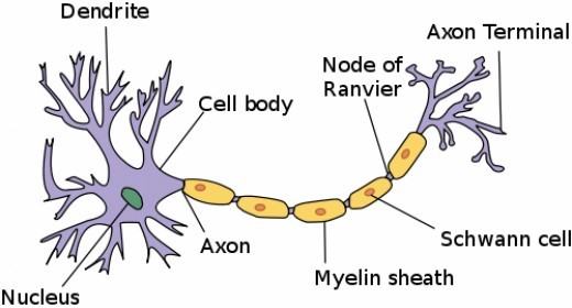
FIGURE 3

ACTION POTENTIAL
FIGURE 4
Figure source- http://www.usdbiology.com/cliff/Courses/General%20Biology/7%20neuronXIII-XIV.html
ACTION POTENTIAL IN NEURON

FIGURE SOURCE-
FIGURE 5
HOW ATP SUPPLIES ENERGY BROKEN BY ATPase
SODIUM POTASSIUM CHANNEL ON CELL WALL

FIGURE 6
Figure source- https://en.wikipedia.org/wiki/ATPase
SYNAPSE/SYNAPTIC CLEFT

FIGURE 7
source- http://cephalove.blogspot.com/2010_05_01_archive.htm
GALVANOMETER TO MESURE ELECTRIC CHARGE DIFFERENCE BETWEEN OUTSIDE CELL WALL AND INSIDE CELL WALL

FIGURE 8
Figure source- http://faculty.washington.edu/chudler/ap.html
ACTION POTENTIAL THE CHARGE DIFFERENCE BETWEEN OUTSIDE CELL WALL AND INSIDE CELL WALL

FIGURE 9
Figure source- https://en.wikipedia.org/wiki/Action_potential
SODIUM ATOM (NA)

Figure -10 source- http://www.chemicalelements.com/elements/na.html
POTASSIUM ATOM (K)

Figure 11
source- http://www.chemicalelements.com/elements/k.html
CALCIUM ATOM (CA)

Figure 12 source- http://www.chemicalelements.com/elements/ca.html
ATP HYDROLYSIS

FIGURE 13
FIGURE LINK-
REFERENCES-
DATE- 11/20/2021
BLOG LINK-
CHAPTER 4, BLOOD SUPPLY OF HEART & CARDIO-PULMONARY RESUSCITATION OR CPR
BLOOD SUPPLY OF HEART & CARDIO-PULMONARY RESUSCITATION OR CPR
[ ইংরেজীতে প্রকাশিত করা হল কিছু কিছু বাংলাদেশী বংশোদ্ভূত বিদেশে অবস্থানরত ছাত্র পাঠকেরা যারা বাংলা ভাষা পড়তে পারেনা, তাদের অনুরোধে, দুখিত ]
Dear viewers
In this chapter we are showing how blood is supplied to the Heart. It has also shown how blood supply in heart is blocked through narrowing of the Coronary artery and what measure (BY-PASS SURGERY) is taken then.
It has also shown if someone’s heart- lungs collapse, how CPR or CARDIO-PULMONARY RESUSCITATION is performed instantly on emergency basis that might save the victim’s life. Such patient cannot be saved if CPR is delayed more than 15 minutes waiting to carry hospital for doctor’s treatment.
A doctor can help nothing to a delayed collapse patient. But an attendant nearby can save such patient’s life by performing CPR instantly.
This is a very preliminary knowledge that might be helpful for the students who like to admit into medical professions.
Please open the YouTube line to understand clearly.
HEART BLOOD SUPPLY
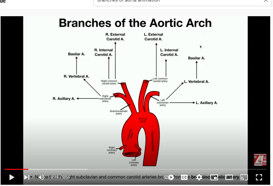
Figure 1
BRANCHES OF THE AORTA (IMAGE)
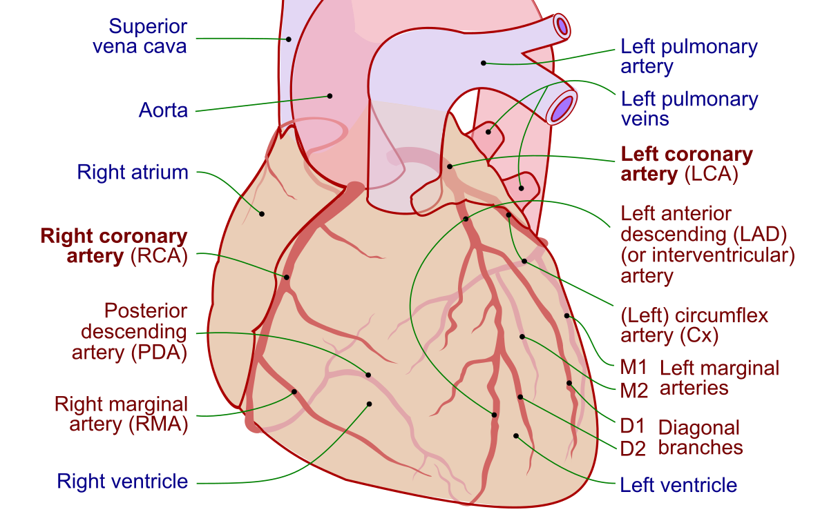
Figure 2
Heart blood supply (image.)
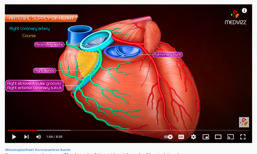
FIGURE 3
LINK- https://www.youtube.com/watch?v=ggj4OWNkBbw
Coronary arteries Anatomy / Blood supply of Heart / Arterial supply of heart: Animation
Coronary Artery Bypass Graft (CABG)
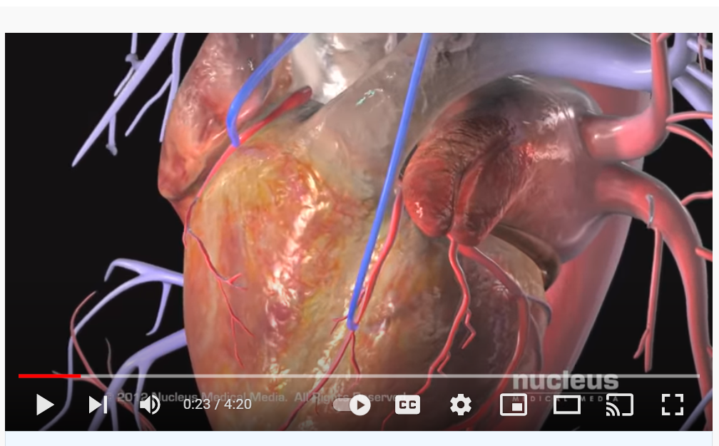
FIGURE 4
LINK- Coronary Artery Bypass Graft (CABG) – YouTube
Coronary Artery Bypass Graft (CABG)
CADIO-PULMONARY RESUSCITATION (CPR)
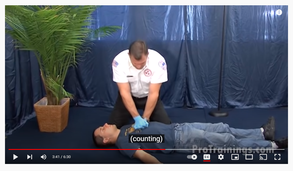
FIGURE 5
Video link- https://www.youtube.com/watch?v=OaSovqEimyA
CPR (CARDIO PULMONARY RESUSCITATION)
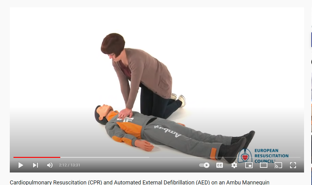
Figure 6
Video link- https://www.youtube.com/watch?v=eNr9x3VJZyM
Date-11/13/2021
Blog link-https://chkdr02.wordpress.com/2021/11/13/blood-supply-of-heart-cardio-pulmonary-resuscitation-or-cpr/
CHAPTER 3. STRUCTURE OF HEART WALL
CHAPTER 3, STRUCTURE OF THE HEART WALL
[ ইংরেজীতে প্রকাশিত করা হল কিছু কিছু বাংলাদেশী বংশোদ্ভূত বিদেশে অবস্থানরত ছাত্র পাঠকেরা যারা বাংলা ভাষা পড়তে পারেনা, তাদের অনুরোধে, দুখিত ]
Dear views,
Dear viewers
In this chapter we are discussing to have a preliminary knowledge about the structure of HEART WALL.
It might be at least a little beneficial for the students having aim to admit medical line.
Please open the links to understand the picture clearly.
IMAGE OF HEART WALL
FIGURE 1

Figure 2
https://www.youtube.com/watch?v=FNsdIuxsrOA
Your Heart Sits In a Sac??? | Pericardium & Pericarditis
LAYERS OF HEART WALL
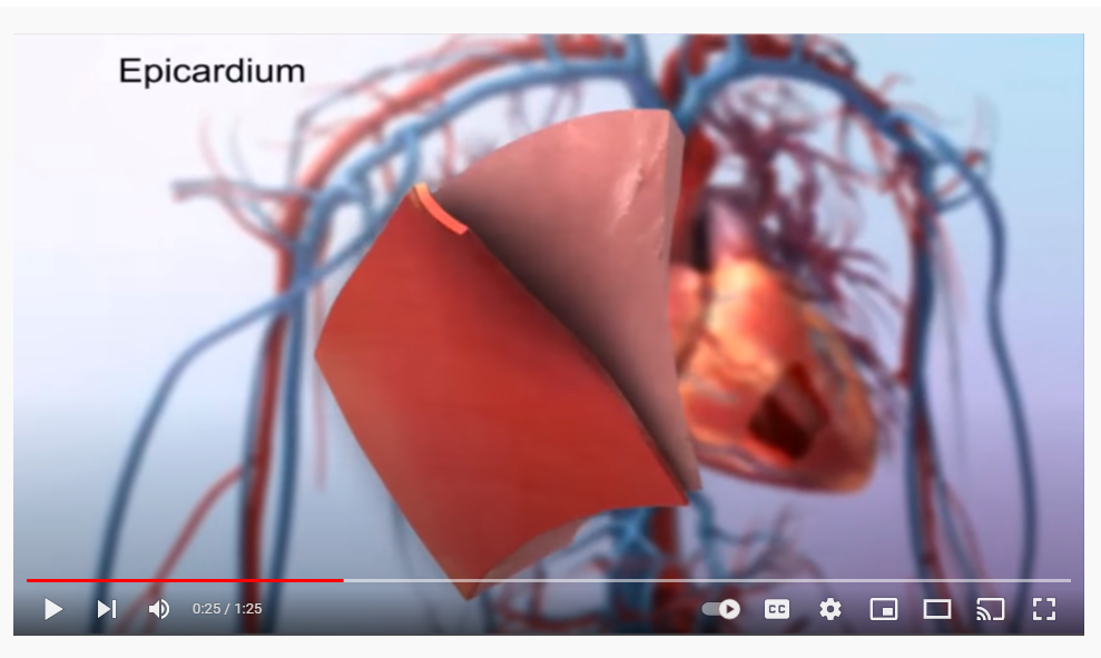
FIGURE 3
https://www.youtube.com/watch?v=9q9sxZTUAd4
