
CHAPTER 53, BLOOD SUPPLY AND NERVE SUPPLY OF EXTERNAL EAR
[ইংরেজীতে প্রকাশিত করা হল কিছু কিছু বাংলাদেশী বংশোদ্ভূত বিদেশে অবস্থানরত ছাত্র পাঠকেরা যারা বাংলা ভাষা পড়তে পারেনা, তাদের অনুরোধে, দুখিত]
BRANCHES OF AORTA
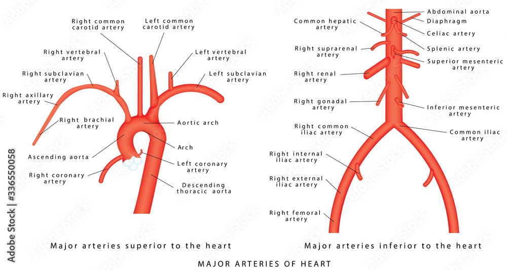
FIGURE- 1
SOURCE OF FIGURE- https://stock.adobe.com/search/images?k=aorta&asset_id=336550058
BRANCHES OF COMMON CAROTID ARTERY
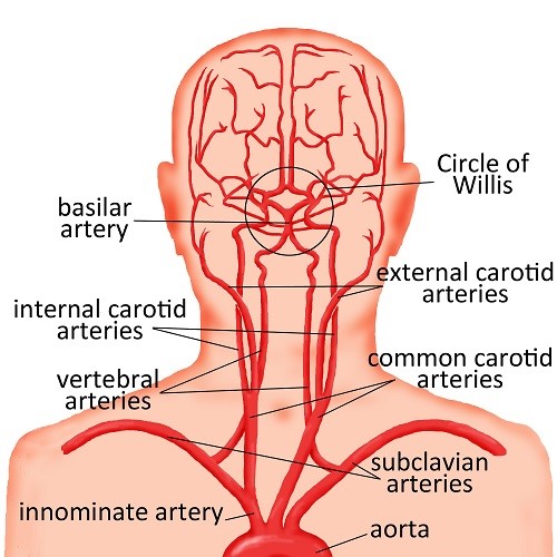
FIGURE-2
SOURCE OF FIGURE- https://www.angiologia.sk/ba/vysetrenia/vysetrenie-krcnych-arterii/?lang=en
BLOOD SUPPLY OF EXTERNAL EAR
FIGURE-
SOURCE OF FIGURE- https://plasticsurgerykey.com/ear/
BLOOD SUPPLY || EXTERNAL EAR ANIMATED SERIES || MEDI CREATOR
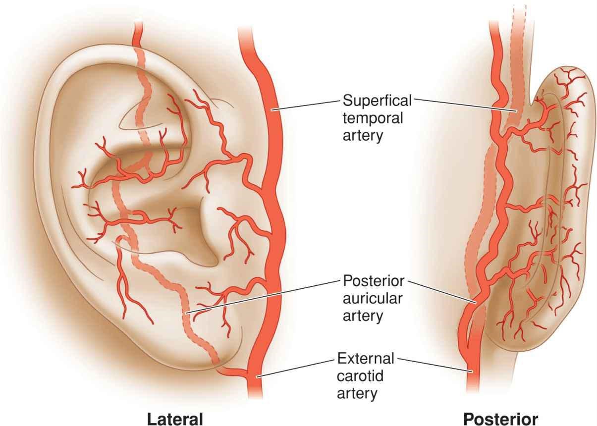
FIGURE-3
SOURCE OF FIGURE- https://www.youtube.com/watch?v=DMMC6oZGQ58
BLOOD SUPPLY #2 || EXTERNAL EAR ANIMATED SERIES || MEDI CREATOR #externalear #bloodsupply
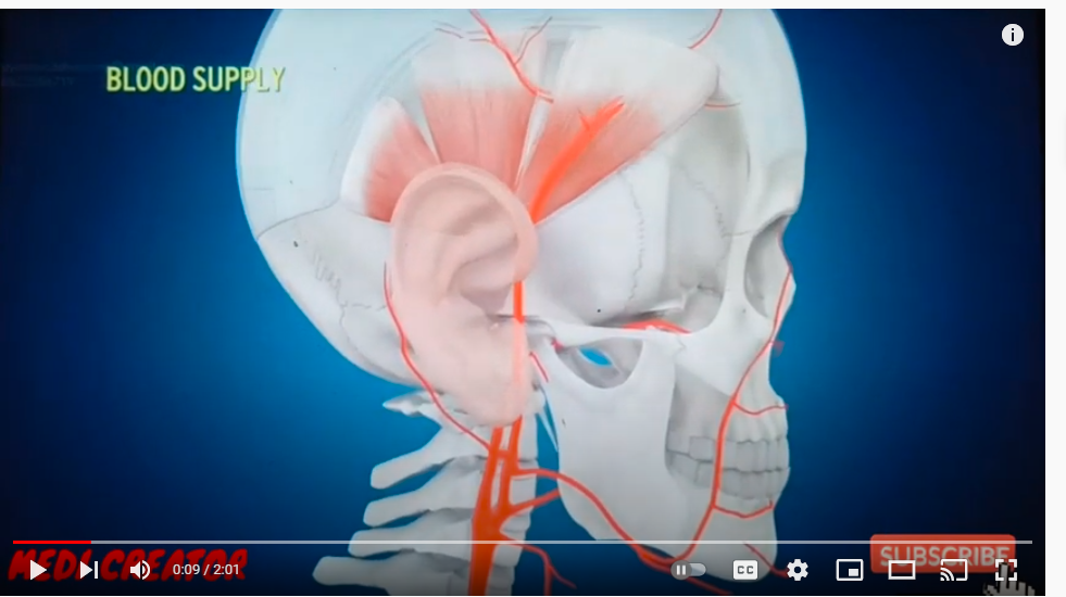
FIGURE-4
SOURCE OF FIGURE- https://www.youtube.com/watch?v=DMMC6oZGQ58
BLOOD SUPPLY #2 || EXTERNAL EAR ANIMATED SERIES || MEDI CREATOR #externalear #bloodsupply
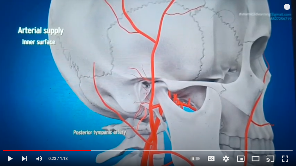
FIGURE 5
SOURCE OF LINK- https://www.youtube.com/watch?v=SBdaNAve3O0
DATE- 10/27/2022
BLOG LINK-
CHAPTER 52, WHAT IS 8TH CRANIAL NERVE? WHAT IS COCHLEAR NERVE? WHAT IS “WERNICKE” AREA AND “BROCA’S” AREA? WHAT IS LIMBIC AREA?
[ইংরেজীতে প্রকাশিত করা হল কিছু কিছু বাংলাদেশী বংশোদ্ভূত বিদেশে অবস্থানরত ছাত্র পাঠকেরা যারা বাংলা ভাষা পড়তে পারেনা, তাদের অনুরোধে, দুখিত]
Dear viewers,
It is written as a primary knowledge for the students who are interested in studying medical sciences. Today we are going to discuss 8th cranial nerve in particular the Cochlear nerve. WHAT IS “WERNICKE” AREA AND “BROCA’S” AREA? WHAT IS LIMBIC AREA?
BASE OF THE SKULL

FIGURE 1
SOURCE OF THE FIGURE- https://bio.libretexts.org/Courses/West_Hills_College_-_Lemoore/Human_Anatomy_Laboratory_Manual_(Hartline)/16%3A_Special_Senses_of_the_Nervous_System/16.03%3A_Auditory_and_Equilibrium_Anatomy
PRIMARY AUDITORY CORTEX, WERNICKE’S AREA OF THE BRAIN
Summary
The area of the associative auditory cortex is called Wernicke’s area. Simply put, this brain segment is responsible for understanding speech. As we will elaborate on brain centers for speech, words, and language in this article, it is important to emphasize the difference between Broca’s area and Wernicke’s area.
Broca’s speech area is in charge of grammatical details and the correct order of words that makes the speech fluent. Damage to this zone leads to disorders of speech in terms of fluency. There are also difficulties in using prepositions, adverbs, and conjunctions.
On the other hand, in 1874, German anatomist Carl Wernicke (1848–1905) found that patients with problems with language comprehension had a part of the left hemisphere of the brain-damaged. This area was named the Wernicke’s area or region. We can say that Wernicke’s area processes the meanings of sounds (1). This zone adds meaning and understanding of the words that we hear.
In this article, we will talk about its anatomical position, relation to other areas, its structure, and its function. Finally, we will provide information on the damage of the Wernicke’s Area, its consequences, and possible treatments.
The Discovery of the Wernicke Area
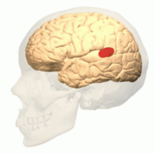
FIGURE-2
In the 19th century, neuroscientists were trying to discover and localize functions and abilities in our brain and relate them to their responsible centers. One of the areas of their interest was language production, processing, and understanding.
Paul Broca was one of the pioneers when it comes to language centers research. In the early 1870s, he made a remarkable discovery. Namely, he identified the brain region responsible for language production.
Paul Broca also made research on patients with problems in producing language, especially in cases when they do understand the language but cannot speak. Broca found that lesions in a specific part of the frontal lobe lead to that very disorder. It is this brain area that we today know as the Broca’s area.
The other key neuroscientist of the 19th century is German neurologist, Carl Wernicke, as mentioned in the introduction. This neurologist was interested in patients with a similar, yet so different problem.
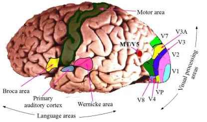
FIGURE-3
SOURCE OF FIGURE AND TEXT- https://human-memory.net/wernickes-area/
WERNICKE’S AREA OF THE BRAIN
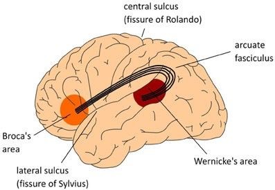
FIGURE-4
SOURCE OF THE LINK- https://human-memory.net/wernickes-area/
CEREBRAL CORTEX NUMBER
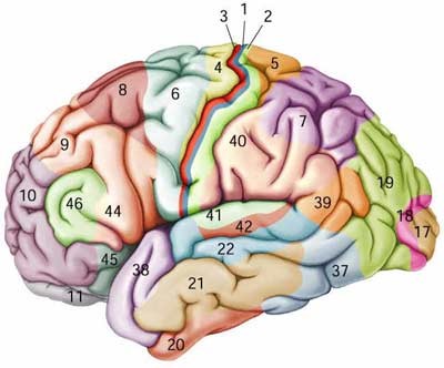
FIGURE- 5
SOURCE OF LINK- https://thebrain.mcgill.ca/flash/capsules/outil_jaune05.html
ANATOMY OF THE BRAIN-
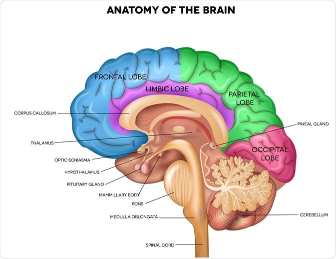
FIGURE-6
SOURCE F THE FIGURE-
https://www.news-medical.net/health/The-Anatomy-of-the-Human-Brain.aspx
BRAIN STEM
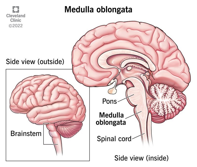
FIGURE-7
SOURCE OF THE FIGURE- https://my.clevelandclinic.org/health/body/23001-medulla-oblongata#anatomy
CEREBRAL COTEX
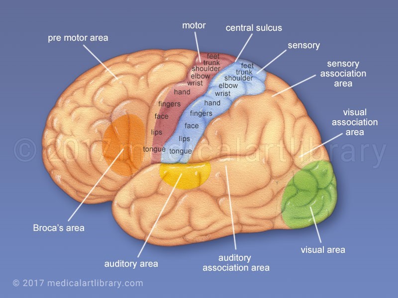
FIGURE- 8
SOURCE OF THE FIGURE- https://medicalartlibrary.com/cerebral-cortex/
Neurology | Vestibulocochlear Nerve | Cranial Nerve VIII: Auditory Pathway
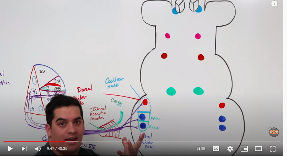
FIGURE- 9
SOURCE OF THE LINK- https://www.youtube.com/watch?v=V8AZ6QygeYs
Auditory Pathway | Easy | Physiology | Primary (lemniscal) pathway
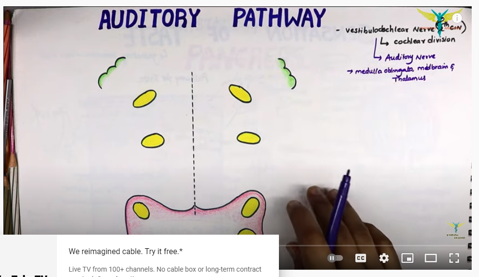
FIGURE- 10
SOURCE OF LINK- https://www.youtube.com/watch?v=J7ZQsK6_Zcc
AUDITORY PATHWAY | ENT |PHYSIOLOGY
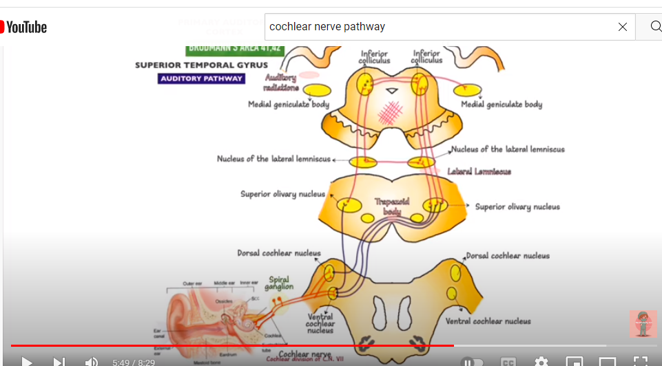
FIGURE- 11
SOURCE OF LINK- https://www.youtube.com/watch?v=vm7RgNvKwMI
The Limbic System
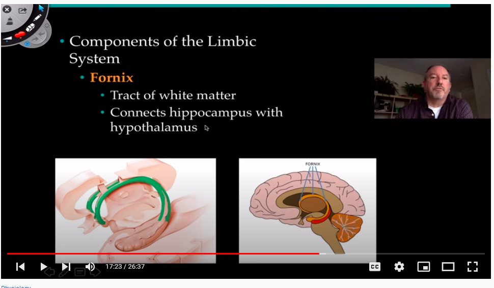
FIGURE 12
SOURCE OF THE LINK- https://www.youtube.com/watch?v=mN9y2LTg8nk&list=LLwbBmvre5US6kA7XCASuXyQ
Date- 10/17/2022
BLOG LINK
CHAPTER 51, WHAT IS 8TH CRANIAL NERVE? WHAT IS VESTBULAR NERVE?
RIDA’S CLASS COMPLETED UPTO CHAPTER 51
[ইংরেজীতে প্রকাশিত করা হল কিছু কিছু বাংলাদেশী বংশোদ্ভূত বিদেশে অবস্থানরত ছাত্র পাঠকেরা যারা বাংলা ভাষা পড়তে পারেনা, তাদের অনুরোধে, দুখিত]
Dear viewers,
It is written as a primary knowledge for the students who are interested in studying medical sciences. Today we are going to discuss 8th cranial nerve in particular the vestibular nerve.
What is 8th cranial nerve? What is vetibular nerve?
Answer- The vestibulocochlear nerve consists of the vestibular and cochlear nerves, also known as cranial nerve eight (CN VIII). Each nerve has distinct nuclei within the brainstem. The vestibular nerve is primarily responsible for maintaining body balance and eye movements, while the cochlear nerve is responsible for hearing
What is the nucleus of cranial nerve?
Answer- A cranial nerve nucleus is a collection of neurons (gray matter) in the brain stem that is associated with one or more of the cranial nerves. Axons carrying information to and from the cranial nerves form a synapse first at these nuclei.
What is brain stem?
Answer- The brain stem is the lower part of the brain that’s connected to the spinal cord (part of the central nervous system in the spinal column). The brain stem is responsible for regulating most of the body’s automatic functions that are essential for life. These include: breathing.
UTRICLE SACULE VESTIVULE

FIGURE-1
SORCE OF THE FIGURE- – https://quizlet.com/315191709/vestibular-equilibrium-cerebellum-diagram/
EAR
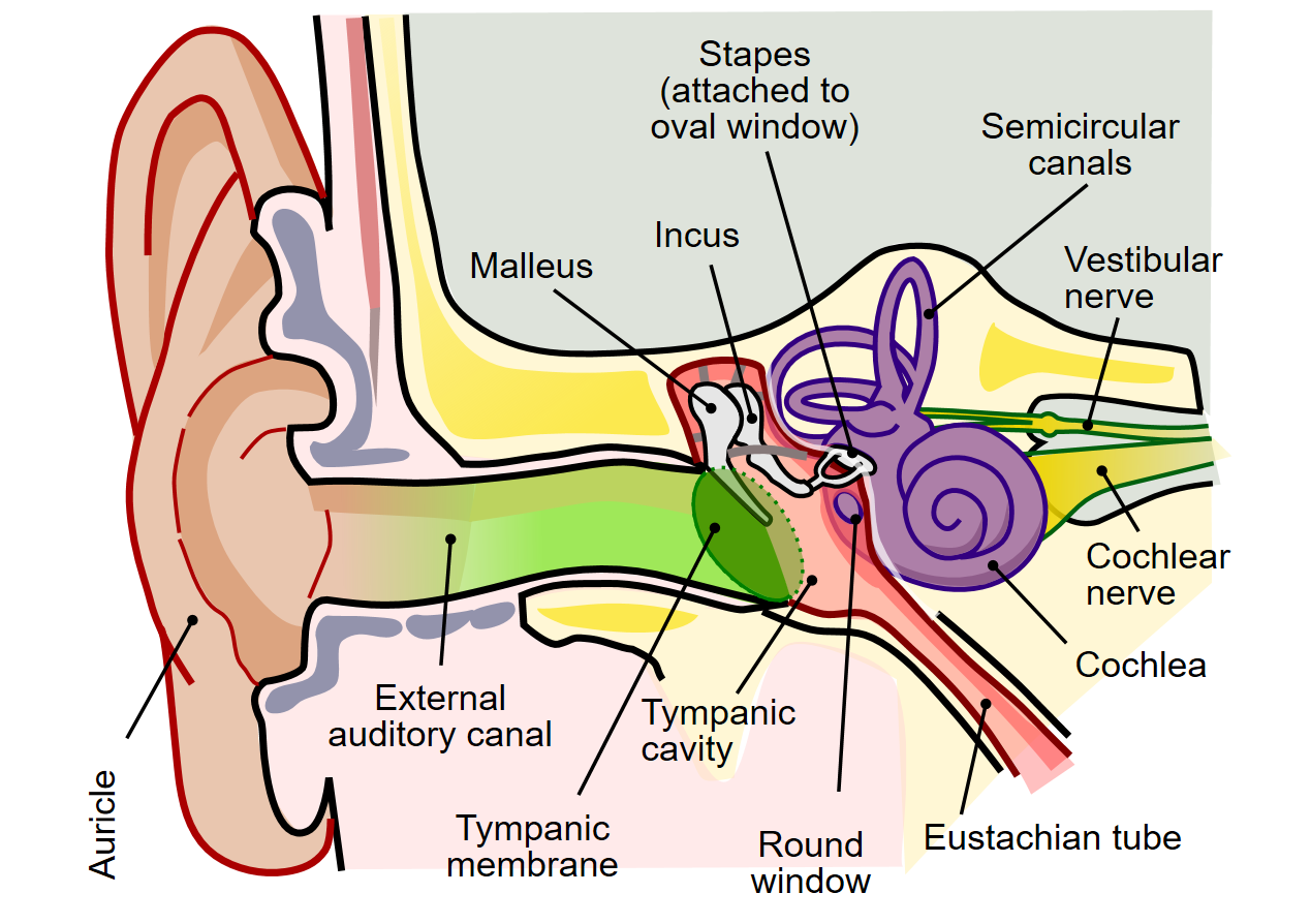
FGURE- 2
SOURCE OF THE FIGURE-
BRAIN STEM

FIGURE-3
SOURCE OF THE FIGURE- https://my.clevelandclinic.org/health/body/23001-medulla-oblongata#anatomy
ANATOMY OF THE BRAIN-

FIGURE-4
SOURCE OF THE FIGURE- https://www.news-medical.net/health/The-Anatomy-of-the-Human-Brain.aspx
CEREBRAL COTEX

FIGURE-5
SOURCE OF THE FIGURE- https://medicalartlibrary.com/cerebral-cortex/
INTERNAL AQUOSTIC MEATUS ON BASE OF SKULL
FIGURE-6
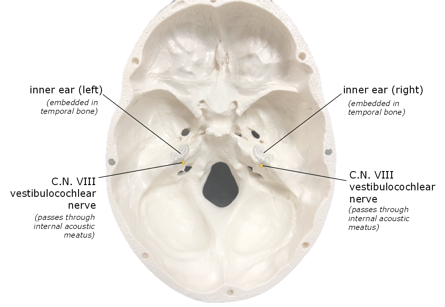
SOURCE OF THE LINK-
Neurology | Vestibulocochlear Nerve | Cranial Nerve VIII: Vestibular Pathway
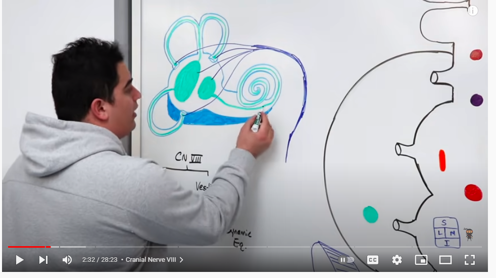
FIGURE-7
SOURCE OF FIGURE- https://www.youtube.com/watch?v=pxga9ci2ets
Date- 10/13/2022
BLOG LINK-
CHAPTER 50, WHAT IS ORGAN OF CORTI IN COCHLEA?
[ইংরেজীতে প্রকাশিত করা হল কিছু কিছু বাংলাদেশী বংশোদ্ভূত বিদেশে অবস্থানরত ছাত্র পাঠকেরা যারা বাংলা ভাষা পড়তে পারেনা, তাদের অনুরোধে, দুখিত]
Dear viewers,
It is written as a primary knowledge for the students who are interested in studying medical sciences. Today we are going to discuss what is organ of corti in cochlea?
WAHAT IS ORGAN OF CORTI?- The Organ of Corti is an organ of the inner ear located within the cochlea which contributes to audition. The Organ of Corti includes three rows of outer hair cells and one row of inner hair cells. Vibrations caused by sound waves bend the stereocilia on these hair cells via an electromechanical force.
Why is it called organ of Corti?
The organ of Corti is named after Italian anatomist Alfonso Corti, who first described it in 1851. Viewed in cross section, the most striking feature of the organ of Corti is the arch, or tunnel, of Corti, formed by two rows of pillar cells, or rods. The pillar cells furnish the major support of this structure.

Figure-1
Source of the figure- https://www.britannica.com/science/ear/Organ-of-Corti
Special Senses | Cochlea | Spiral Organ of Corti
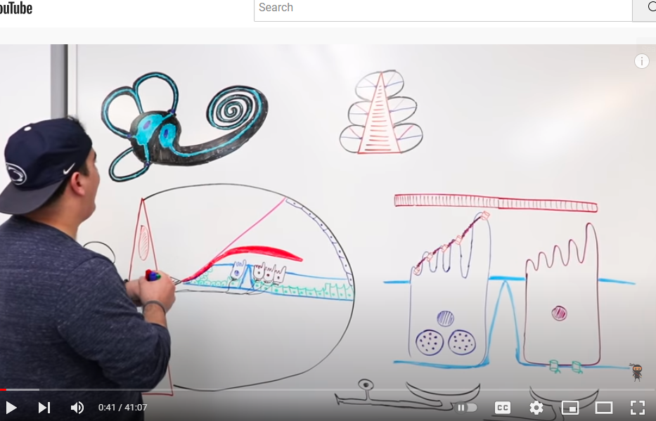
FIGURE-2
Source of link- Special Senses | Cochlea | Spiral Organ of Corti – YouTube
Ear Organ of Corti (Full Version)

FIGURE NO-3
SOURCE OF YOU TUBE LINK- https://www.youtube.com/watch?v=1JE8WduJKV4&list=LLwbBmvre5US6kA7XCASuXyQ&index=2&t=107s
ORGAN 0F CORTI COCHLEA

FIGURE-4
YOU TUBE LINK- The Organ of Corti A Simple Explanation – YouTube
Date- 10/05/2022
BLOG LINK-
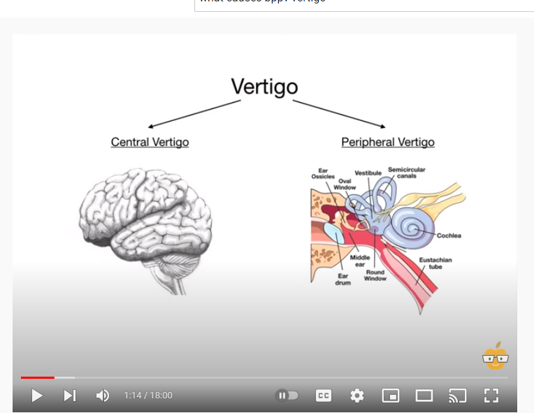
CHAPTER 49, WHAT IS OTOCONIA? WHAT IS VERTIGO? WHAT IS NYSTAGMUS?
[ইংরেজীতে প্রকাশিত করা হল কিছু কিছু বাংলাদেশী বংশোদ্ভূত বিদেশে অবস্থানরত ছাত্র পাঠকেরা যারা বাংলা ভাষা পড়তে পারেনা, তাদের অনুরোধে, দুখিত]
Dear viewers,
It is written as a primary knowledge for the students who are interested in studying medical sciences. Today we are going to discuss what is Otoconia? What is Vertigo? What is Nystagmus?
What is otoconia?
BPPV occurs when tiny calcium crystals called otoconia come loose from their normal location on the utricle, a sensory organ in the inner ear. If the crystals become detached, they can flow freely in the fluid-filled spaces of the inner ear, including the semicircular canals (SCC) that sense the rotation of the head.
Source – Google.
Benign Paroxysmal Positional Vertigo (BPPV)
Text link-
Special Senses | Semicircular Canals | Cristae Ampullaris | BPPV
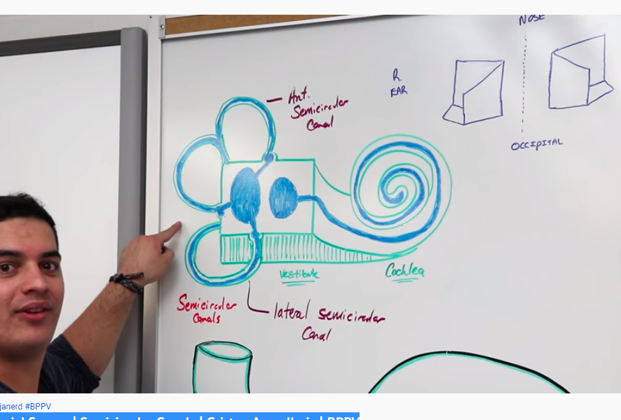
Figure-1
Special Senses | Semicircular Canals | Cristae Ampullaris | BPPV – YouTube
Vertigo (Different Types, Dix-Hallpike Maneuver, Treatment)
FIGURE—2
YOU TUBE LINK- https://www.youtube.com/watch?v=cbGmR4v3bkU
What Is Benign Paroxysmal Positional Vertigo?

FIGURE-3
YOU TUBE LINK- https://www.youtube.com/watch?v=1AfvNsaQnTE
Nystagmus Eyes Explained | Involuntary Repetitive Eye Movement

FIGURE-4
YOU TUBE LINK- https://www.youtube.com/watch?v=CgaKef1cnTw
DATE: 9/08/2022
Please look the blog post-
DATE: 9/15/2022
Please look the blog post-
CHAPTER 48, WHAT IS SEMICIRCULAR CANAL? WHAT IS IT’S FUNCTION?
[ইংরেজীতে প্রকাশিত করা হল কিছু কিছু বাংলাদেশী বংশোদ্ভূত বিদেশে অবস্থানরত ছাত্র পাঠকেরা যারা বাংলা ভাষা পড়তে পারেনা, তাদের অনুরোধে, দুখিত]
Dear viewers,
It is written as a primary knowledge for the students who are interested in studying medical sciences. Today we are going to discuss what is semicircular canal, what is it’s function?
Special Senses | Semicircular Canals | Cristae Ampullaris | BPPV

Figure-1
Special Senses | Semicircular Canals | Cristae Ampullaris | BPPV – YouTube
DATE: 9/08/2022
Please look the blog post-
CHAPTER 47, WHAT IS VESTIBULE, MACULAE, UTRICLE & SACULE, HOW BODY BALANCE IS MAINTAINED?
[ইংরেজীতে প্রকাশিত করা হল কিছু কিছু বাংলাদেশী বংশোদ্ভূত বিদেশে অবস্থানরত ছাত্র পাঠকেরা যারা বাংলা ভাষা পড়তে পারেনা, তাদের অনুরোধে, দুখিত]
Dear viewers,
It is written as a primary knowledge for the students who are interested in studying medical sciences. Today we are discussing how body balance is maintained?
Special Senses | Vestibule | Maculae: Utricle & Saccule
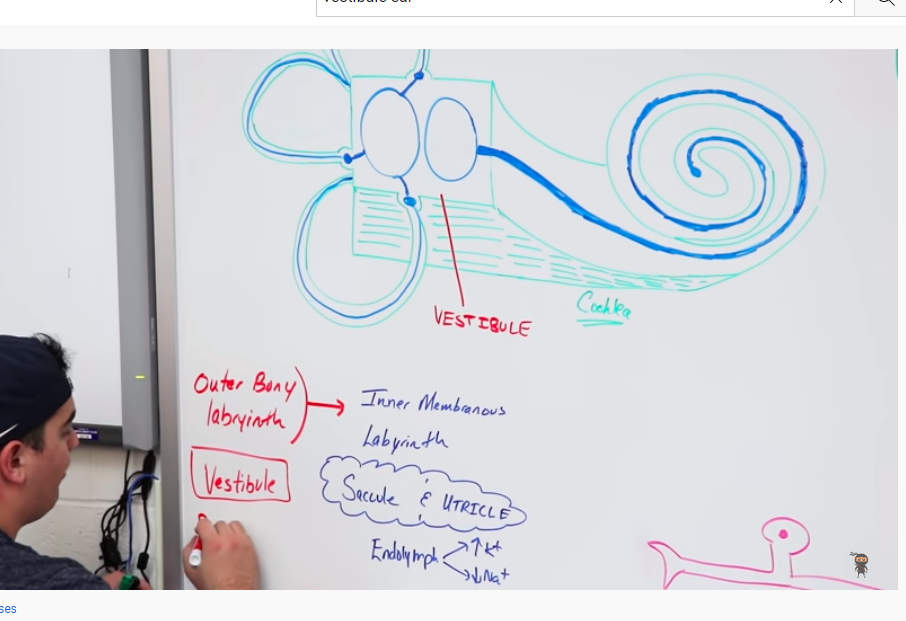
FIGURE-1
SOURCE OF THE YOU TUBE LINK-
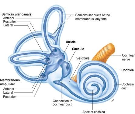
FIGURE 2
SOURCE OF LINK- https://quizlet.com/315191709/vestibular-equilibrium-cerebellum-diagram/
Date-8/31/2022
Link of the blog-
CHAPTER 46, WHAT IS ANATOMY OF COCHLEA AND HOW IT WORKS? WHAT IS ACTION POTENTIAL?WHAT IS GANGLION? WHAT IS NEUROTRANSMITTER?
Dear viewers,
[ইংরেজীতে প্রকাশিত করা হল কিছু কিছু বাংলাদেশী বংশোদ্ভূত বিদেশে অবস্থানরত ছাত্র পাঠকেরা যারা বাংলা ভাষা পড়তে পারেনা, তাদের অনুরোধে, দুখিত]
It is written as a primary knowledge for the students who are interested in studying medical sciences. Today we are going to discuss the anatomy of cochlea and how it works.What is action potential? what is ganglion? What is neurotransmitter?
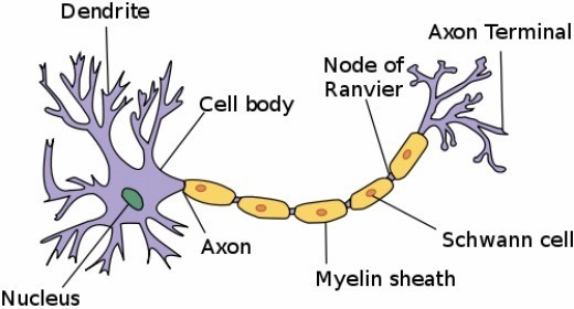
Figure-1
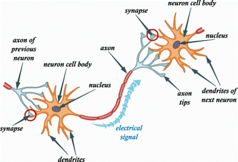
Figure 2
source- http://www.usdbiology.com/cliff/Courses/General%20Biology/7%20neuronXIII-XIV.html
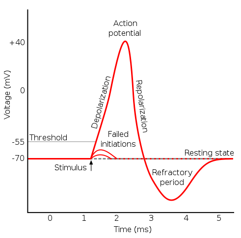
Figure-3
Figure-4
Link of this figure-
https://en.wikipedia.org/wiki/File:Action_Potential.gif
source of the figures- WHAT IS ACTION POTENTIAL (1)
BLOG LINK https://chkdr02.wordpress.com/2021/11/20/what-is-action/
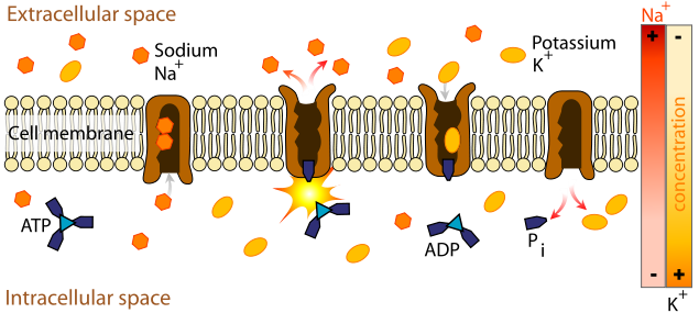
Figure- 5
Source of link- https://en.wikipedia.org/wiki/ATPaseSYNAPSE/SYNAPTIC CLEFT

FIGURE 6
source- http://cephalove.blogspot.com/2010_05_01_archive.htm
–
WHAT IS ACTION POTENTIAL?
SEE THE LINK- What Is DNA? Chapter 17th, The Nobel Laureates In Physiology Or Medicine In 2013, What Is Neuron (2)
https://dnaandthemystryofhumanbody.wordpress.com/wp-admin/post.php?post=172&action=edit&calypsoify=1
Special Senses | Cochlea | Spiral Organ of Corti

Figure- figure 7
Source of you tube- Special Senses | Cochlea | Spiral Organ of Corti – YouTube
Special Senses | Vestibule | Maculae: Utricle & Saccule
DATE: 8/25/2022
Please look the blog post-
